Kinesin Motility Assay Biochem Kit (fluorescence format)
Important Notice:
This product is no longer available. Individual components may still be purchased (see links in kit contents below).
Product Uses Include
- To determine whether a protein or compound alters kinesin or dynein-microtubule mobility.
- To test other proteins for microtubule mobility.
Introduction
This is a Fluorescence based motility assay. To perform this assay effectively you will require a video microscope system with a shutter mechanism for time lapse image acquisition. Kinesin or dynein motors are coupled to the surface of a mcroscope perfusion chamber (kinesin heavy chain motor domain is included in the kit as a positive control) and fluorescent microtubules are added. The motor-induced motility of the microtubules can be monitored and measured in a fluorescence microscope.
Kit contents
The kit contains sufficient materials for 25 assays. The following components are included:
- Tubulin, >99% pure, lyophilized (Cat. # TL238)
- Rhodamine tubulin, lyophilized (Cat. # TL331M)
- Kinesin heavy chain motor domain, lyophilized (positive control) (Cat. # KR01)
- Antifade
- ATP (Cat. # BSA04)
- Microtubule polymerization buffer
- Tubulin glycerol buffer
- Microtubule resuspension buffer
- Blocking solution
- Chamber wash buffer
- Motility buffer
- Paclitaxel (Cat. # TXD01)
- DMSO
- 25 acid washed perfusion chambers
- Chamber filters
- Manual with detailed protocols and extensive troubleshooting guide
Equipment needed
- Fluorescence microscope equipped with filters to detect rhodamine and ready for time-lapse image capturing
Example results
The motility of microtubules in a kinesin heavy chain (Cat. # KR01) coated chamber was followed with time lapse microscopy (Fig. 1)
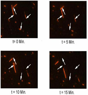
Figure 1. Microtubule Motility Assay. The field was selected to show a sparse area of MT coverage in order to allow clear visualization of MT motility. Each frame represents a 5 min time-point, from 0 to 15 min. The position of each MT at time zero is marked by an arrow. The arrow position remains identical in each frame to serve as a reference point for MT movement. To obtain an average MT velocity, we recommend measure motility of approximately 30 - 50 individual MTs.
For product Datasheets and MSDSs please click on the PDF links below. For additional information, click on the FAQs tab above or contact our Technical Support department at tservice@cytoskeleton.com
If you have any questions concerning this product, please contact our Technical Service department at tservice@cytoskeleton.com
Question 1: At what speed does the kinesin motor move in this assay?
Answer 1: The recombinant kinesin motor provided with this motility assay moves very slowly (average of 0.5 μm/min) and motility will not be seen without the use of a time lapse video microscopy system set on 2 to 5 min per frame. Native or full length recombinant kinesin moves with a velocity of 30 - 40 μm/min.
Question 2: I have high fluorescent background. How can I reduce it?
Answer 2: There will be some noticeable background fluorescence. The background fluorescence results from unpolymerized tubulin and can be removed by passing the microtubules over a glycerol cushion (see the manual for a detailed protocol).
If you have any questions concerning this product, please contact our Technical Service department at tservice@cytoskeleton.com

