Fibronectin proteins and derivatives
Unlock the full potential of your extracellular matrix (ECM) and cell adhesion studies with our advanced line of fibronectin protein products.
A broad selection of fluorescent derivatives spanning a wide spectral range.
Biotinylated with a long carbon chain spacer to enhance biotin accessibility for coupling.
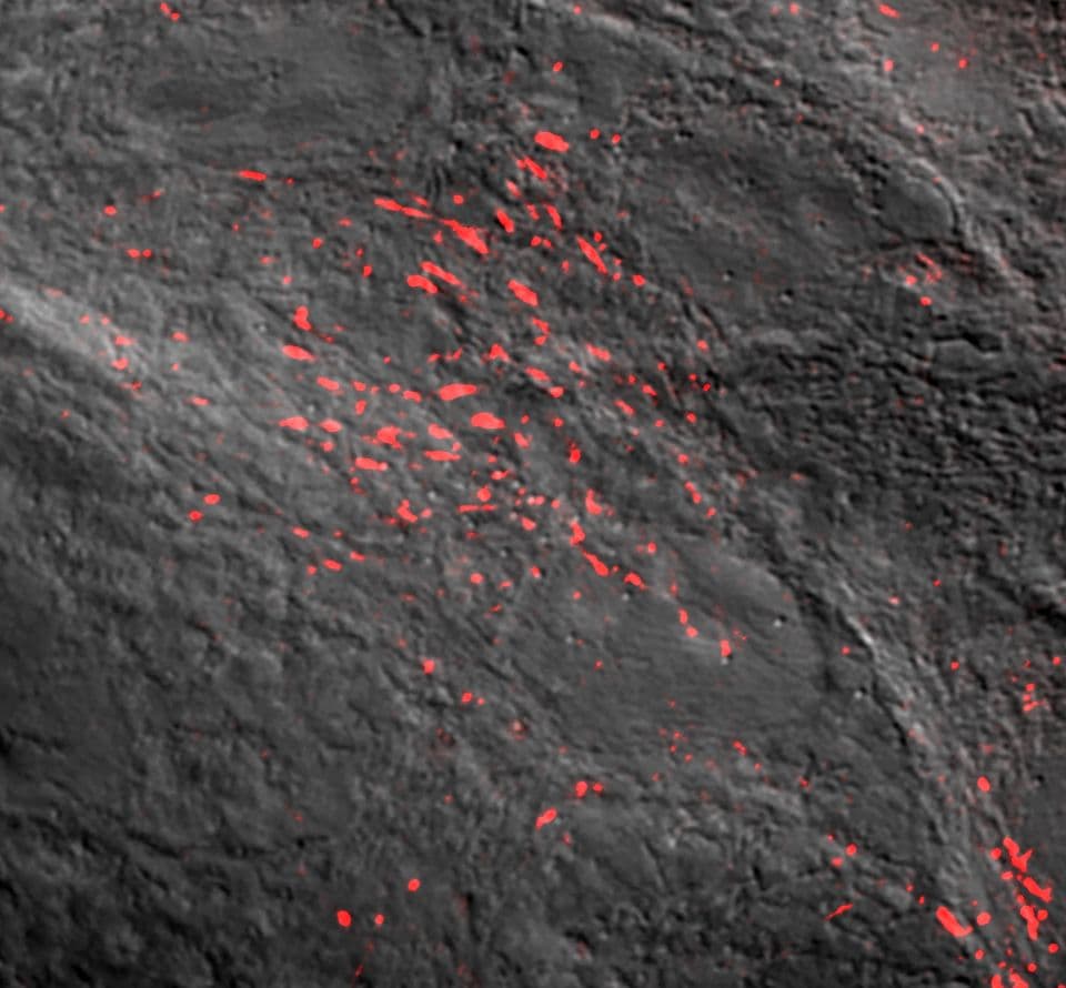
Validation of bioactivity for real-time imaging of fibronectin dynamics.
Excellent for cell adhesion assays, live-cell imaging, and ECM remodeling studies.
Designed for demanding academic and biotech research, our fibronectin reagents support reproducible, high-quality results across a range of applications.
Unlock the full potential of your extracellular matrix (ECM) and cell adhesion studies with our advanced line of fibronectin protein products.
A broad selection of fluorescent derivatives spanning a wide spectral range.
Biotinylated with a long carbon chain spacer to enhance biotin accessibility for coupling.
Validation of bioactivity for real-time imaging of fibronectin dynamics.
Excellent for cell adhesion assays, live-cell imaging, and ECM remodeling studies.
Designed for demanding academic and biotech research, our fibronectin reagents support reproducible, high-quality results across a range of applications.