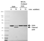
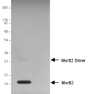
MICAL-1 Protein 6xHis

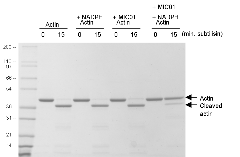
MICAL (MIC01) is an intracellular flavoprotein monoxygenase, conserved from insects to mammals, that functions as a catalyst for oxidation-reduction (redox) reactions. Terman’s group showed that MICAL interacts with F-actin and uses NADPH as a cofactor to oxidize actin at Met44 and Met47. As part of the MOXtrue™ product line, Human MICAL1 protein (MIC01) [accession # Q8TDZ2 (MICA1_HUMAN)] has been produced and purified from a bacterial expression system. The recombinant protein is N-terminally 6x Histidine tagged and is comprised of the Redox and CH domains (aa: 1-612) of the full length protein. Validation studies have shown that MIC01 can oxidize actin which will reduce subtilisin A cleavage by >90% in limited proteolysis assays. MIC01 has also been validated for the production of oxidized actin which can be used in downstream applications such as sedimentation assays and actin polymerization assays.
To learn more about using MICAL as a research tool see our Datasheet
To learn more about the MICAL/MsrB/Actin physiological redox system see our Newsletter
Each lot of purified protein is quality controlled to provide high batch to batch consistency, see COA documents.
Validated Applications
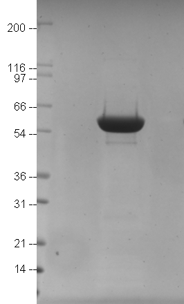
MICAL-1 (MIC01) Protein Purity Determination A 10 μg sample of MICAL-1 protein was sepa-rated by electrophoresis in a 4- 20% Tris-Glycine gel and stained with Coomassie Blue. Protein quantitation was performed using the Precision RedTM Protein Assay Reagent (Cat.# ADV02). Mark12 standard molecular weight markers are from Invitrogen. |
Subtilisin Assay on MsrB2 Treated Oxidized Actin MICAL-oxidized actin (MetO Actin) (Cat# MXA95) was diluted to 0.1 mg/ml (2.3 μM) and left untreated, or treated with DTT (10 mM), MB201 (0.3 μM) , or a combination of both (see method). 2 μg of each sample was then left untreated, or treated with subtilisin (1:200 w/w) for 15 min. Samples were then separated by SDS-PAGE and visualized with Coomassie staining. Click here for a detailed method |
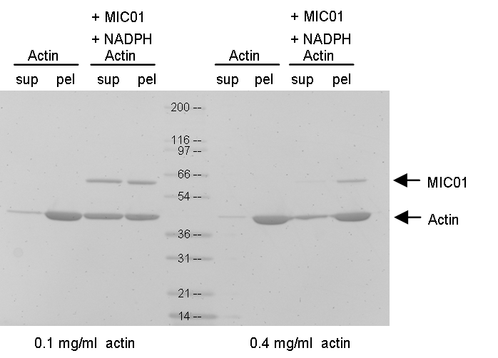
Actin Sedimentation +/- MICAL-1 Treatment
Actin (Cat# AKL99) was diluted to 0.2 mg/ml (4.6 μM) or 0.8 mg/ml (18.4 μM) (see method). Samples were then incubated with 2x polymerization buffer or 2x polymerization buffer supplemented with MIC01 + NADPH for 1 h at room temperatire. Samples were spun in an ultracentrifuge at 100,000 g for 1.5 h. Samples were then separated by SDS-PAGE and visualized with Coomassie staining.
Click here for a detailed method
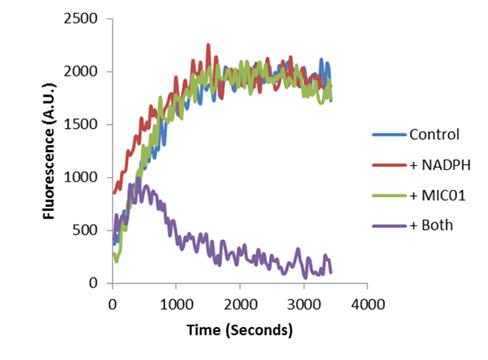
Actin Polymerization +/- MICAL-1 Treatment.
Pyrene labeled-actin (Cat. # AP05) was diluted to 0.1 mg/ml (2.3 μM) (see method). Samples were then incubated with 2x polymerization buffer supplemented with nothing (labeled Control), NADPH alone (labeled +NADPH), MIC01 alone (labeled +MIC01), or the combination of both (labeled +Both). Upon actin polymerization fluorescence was detected with a spectrophotometer (see method below). A.U. = arbitrary units
Click here for a detailed method
For more information contact: signalseeker@cytoskeleton.com
Associated Products:
MOXtrue™ 6xHis MsrB2 Protein (Cat. # MB201)
MOXtrue™ MICAL-oxidized Actin (Cat. # MXA95)
MOXtrue™ MICAL-oxidized Pyrene Labeled Actin (Cat. # MPAX1)
Rabbit Skeletal Muscle Actin (Cat. # AKL95)
Pyrene Labeled Rabbit Skeletal Muscle Actin (Cat.# AP05)
For product Datasheets and MSDSs please click on the PDF links below.
Certificate of Analysis: Lot001
If you have any questions concerning this product, please contact our Technical Service department at tservice@cytoskeleton.com
Visit our Signal-Seeker™ Tech Tips and FAQs page for technical tips and frequently asked questions regarding this and other Signal-Seeker™ products click here
If you have any questions concerning this product, please contact our Technical Service department at tservice@cytoskeleton.coma


