Cdc42 G-LISA GTPase Activation Assay (Colorimetric format) - 24 assays

G-LISA Cdc42 Activation Assay Biochem Kit (Colorimetric format)

$0.00

Product Uses Include

  • Rho signaling pathway studies
  • Rho activation assays with primary cells
  • Rho activation assays with limited material
  • High throughput screens for Rho activation

Introduction
The G-LISA® series of Small G-Protein Activation Assays are ELISA based assays with which you can measure the GTP form of small G-proteins from lysates of cells or tissues and all in less than 3 h. The Cdc42 G-LISA Activation Assay measures the entire level of GTP-loaded Cdc42 protein in cell lysates. The level of activation is measured by reading at OD490nm. For a more detailed introduction on G-LISA assays and a listing of other available G-LISA kits, see our main G-LISA page. For a kit to measure RhoA activation please check webpage BK124, for Rac1 go to BK128 for Rac1,2,3 go to BK125, for RalA go to BK129.

The Cdc42 G-LISA Activation Assay is very sensitive and has excellent accuracy for duplicate samples. See this section on our G-LISA information resource page.


Example results
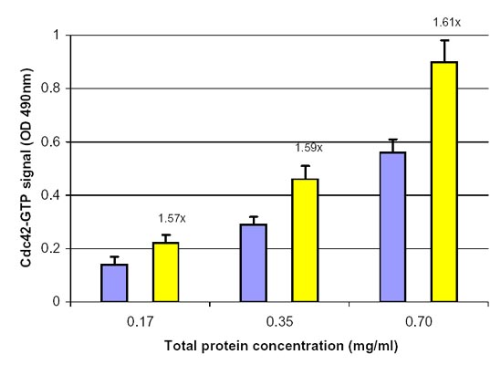
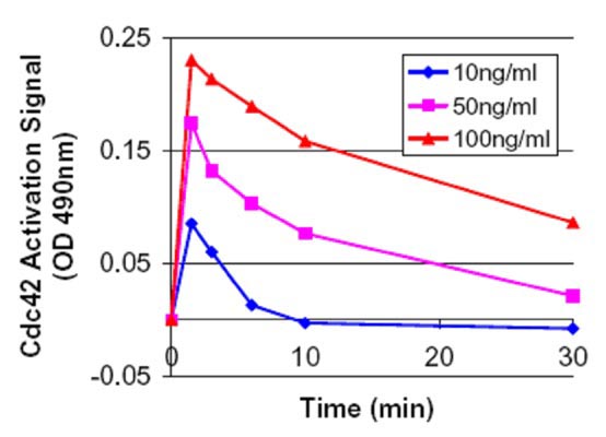
Serum starved Swiss 3T3 cells were stimulated with the Cdc42 activating compound EGF and Cdc42 activation was measured with the G-LISA method (Fig 1 and 2).
BK127_linearity

Figure 1. Cdc42 activation by EGF measured by the Cdc42 G-LISA Activation Assay.  Swiss 3T3 cells were serum starved (SS) for 16 h at 1% serum and 8 h with 0% serum and treated with EGF (100 ng/ml for 2 min). Cell lysates (8, 17, 35 µg) were subjected to the G-LISA assay. Data was read at 490 nm.  Numbers on top the yellow columns indicate the fold increase in signal caused by EGF activation, you will notice the ratio remains the same with different protein loadings indicating good linearity with different protein loadings. 500 µg of the same lysates were subjected to the traditional PAK pull-down assay (Cat.# BK034) with similar results.
BK127_time_course

Figure 2. Time course of Cdc42 activation using EGF at 10, 50 and 100ng/ml .  Swiss 3T3 cells were serum starved (SS) for 16 h at 1% serum and 8 h with 0% serum and treated with EGF (10, 50 and 100 ng/ml for1.5, 3.0, 6.0, 10 and 30 min). Cell lysates subjected to the G-LISA™ assay and OD was read at 490 nm. The “controlled state” serum starved value (0.22) was subtracted from these samples prior to plotting. At 100 ng/ml the total activation was 2.1 fold or 110% over the controlled state at 1.5 min. This type of analysis can be performed in one afternoon.

Kit contents
The kit contains sufficient reagents to perform 24 Cdc42 activation assays. Since the Cdc42-GTP affinity wells are supplied as strips and the strips can be broken into smaller pieces, each kit can be used for anywhere from 1 to 96 assays. The following components are included in the kit:

  1. Cdc42-GTP affinity wells (3 strips of 8 wells each)
  2. Lysis buffer
  3. Antigen presenting buffer
  4. Wash buffer
  5. Antibody dilution buffer
  6. Anti-Cdc42 antibody (Cat.# ACD03)
  7. HRP-labeled secondary antibody
  8. Positive control Cdc42 protein
  9. Protease inhibitor cocktail (Cat. # PIC02)
  10. Color development reagents
  11. Precision Red™ Advanced protein assay reagent (Cat. # ADV02)
  12. Manual with detailed protocols and extensive troubleshooting guide

Equipment needed

    1. 96-well plate spectrophotometer capable of reading 490 nm wavelength
    2. Multichannel or multidispensing pipettor (see Biohit pipettors)
    3. Two orbital microplate shakers capable of 400 rpm shaking (200 to 400 rpm is the possible range)
      Examples are:

Model # 4625  Titer Plate Shaker,  Lab-Line Instruments, Barnstead Intl.(average priced)

Model # RF7854  Digital Microplate Shaker, ML Market Lab,  researchml.com (economical priced)

Model # RF7855  Incubating Microplate Shaker, ML Market Lab, researchml.com (deluxe model)


Go to main G-LISA page

G-LISA Products:
Rac1,2,3 G-LISA Activation Assay, colorimetric format (Cat.# BK125)
Rac1 G-LISA Activation Assay, luminescence format (Cat.# BK126)
RhoA G-LISA Activation Assay, colorimetric format (Cat.# BK124)
RhoA G-LISA Activation Assay, luminescence format (Cat.# BK121)

Associated Products:
Anti-Cdc42 monoclonal antibody (Cat.# ACD03)
Anti-Rac1 monoclonal antibody (Cat.# ARC03)
Anti-RhoA monoclonal antibody (Cat.# ARH03)

G-LISA is a registered trademark of Cytoskeleton, Inc (CO). All rights reserved.

For product Datasheets and MSDSs please click on the PDF links below.   


    AuthorTitleJournalYearArticle Link
    Ji, Juanjuan et al.TMEM16A enhances the activity of the Cdc42-NWASP signaling pathway to promote invasion and metastasis in oral squamous cell carcinomaOral Surgery, Oral Medicine, Oral Pathology and Oral Radiology2024
    Szigeti, Kinga et al.CHRFAM7A diversifies human immune adaption through Ca2+ signalling and actin cytoskeleton reorganizationeBioMedicine2024
    Mendoza-Soto, Pablo et al.Pharmacological Blockade of the Adenosine A2B Receptor Is Protective of Proteinuria in Diabetic Rats, through Affecting Focal Adhesion Kinase Activation and the Adhesion Dynamics of PodocytesCells2024
    Wu, Fei et al.Single-cell analysis identifies conserved features of immune dysfunction in simulated microgravity and spaceflightNature Communications2024
    Lee, Choong Jae et al.The dysadherin/FAK axis promotes individual cell migration in colon cancerInternational Journal of Biological Sciences2024
    Liang, Bishan et al.Cdc42-driven endosomal cholesterol transport promotes collateral resistance in HER2-positive gastric cancerCancer Letters2024
    Adhicary, Subhodip et al.Rbpj Deficiency Disrupts Vascular Remodeling via Abnormal Apelin and Cdc42 (Cell Division Cycle 42) Activity in Brain Arteriovenous MalformationStroke2023
    Szigeti, Kinga et al.Neuronal actin cytoskeleton gain of function in the human braineBioMedicine2023
    Xie, Wangkai et al.ASAP1 activates the IQGAP1/CDC42 pathway to promote tumor progression and chemotherapy resistance in gastric cancerCell Death & Disease2023
    Punessen, Noelle C. et al.A novel anti-apoptotic role for Cdc42/ACK-1 signaling in neuronsMolecular and Cellular Neuroscience2023
    St. Paul, Amanda et al.FXR1 regulates vascular smooth muscle cell cytoskeleton, VSMC contractility, and blood pressure by multiple mechanismsCell reports2023
    Castillo-Azofeifa, David et al.A DLG1-ARHGAP31-CDC42 axis is essential for the intestinal stem cell response to fluctuating niche Wnt signalingCell stem cell2023
    Crespo, Grace Velez et al.The Rac inhibitor HV-107 as a potential therapeutic for metastatic breast cancerMolecular Medicine2023
    Adhicary, Subhodip et al.Rbpj Deficiency Disrupts Vascular Remodeling via Abnormal Apelin and Cdc42 (Cell Division Cycle 42) Activity in Brain Arteriovenous MalformationStroke2023
    Zi, Jingjing et al.PFN1 Inhibits Myogenesis of Bovine Myoblast Cells via Cdc42-PAK/JNKCells2022
    Meng, Zhipeng et al.The Hippo pathway mediates Semaphorin signalingScience Advances2022
    Jiang, Ruiwei et al.CDC42 governs normal oviduct multiciliogenesis through activating AKT to ensure timely embryo transportCell Death & Disease2022
    Ma, Yuanyuan et al.Ror2-mediated non-canonical Wnt signaling regulates Cdc42 and cell proliferation during tooth root developmentDevelopment (Cambridge)2021
    Jozic, Ivan et al.Glucocorticoid-mediated induction of caveolin-1 disrupts cytoskeletal organization, inhibits cell migration and re-epithelialization of non-healing woundsCommunications Biology2021
    Vallejo, Daniela et al.Wnt5a modulates dendritic spine dynamics through the regulation of Cofilin via small Rho GTPase activity in hippocampal neuronsJournal of Neurochemistry2021
    Wurzer, Hannah et al.Intrinsic Resistance of Chronic Lymphocytic Leukemia Cells to NK Cell-Mediated Lysis Can Be Overcome In Vitro by Pharmacological Inhibition of Cdc42-Induced Actin Cytoskeleton RemodelingFrontiers in Immunology2021
    Merlin, Johanna et al.Non-canonical glutamine transamination sustains efferocytosis by coupling redox buffering to oxidative phosphorylationNature Metabolism2021
    Lee, DanielmiR‑769‑5p is associated with prostate cancer recurrence and modulates proliferation and apoptosis of cancer cellsExperimental and Therapeutic Medicine2021
    Salameh, Joëlle et al.Cdc42 and its BORG2 and BORG3 effectors control the subcellular localization of septins between actin stress fibers and microtubulesCurrent Biology2021
    Talamás-Lara, Daniel et al.Entamoeba histolytica and Entamoeba dispar: Morphological and Behavioral Differences Induced by Fibronectin through GTPases Activation and Actin-Binding ProteinsThe Journal of eukaryotic microbiology2020
    Krueger, Irena et al.Reelin amplifies glycoprotein VI activation and alphaiib beta3 integrin outside-in signaling via PLC Gamma 2 and Rho GTPasesArteriosclerosis, Thrombosis, and Vascular Biology2020
    Rong, Zhouyi et al.Activation of FAK/Rac1/Cdc42-GTPase signaling ameliorates impaired microglial migration response to Aβ42 in triggering receptor expressed on myeloid cells 2 loss-of-function murine modelsFASEB Journal2020
    Andrews, Madeline G. et al.Mtor signaling regulates the morphology and migration of outer radial glia in developing human cortexeLife2020
    Choraghe, Rohan P. et al.RHOA-mediated mechanical force generation through Dectin-1Journal of cell science2020
    Malek, Natalia et al.Knockout of ACTB and ACTG1 with CRISPR/Cas9(D10A) technique shows that non-muscle β and γ actin are not equal in relation to human melanoma cells’ motility and focal adhesion formationInternational Journal of Molecular Sciences2020
    Wilson, Kitchener D. et al.Endogenous Retrovirus-Derived lncRNA BANCR Promotes Cardiomyocyte Migration in Humans and Non-human PrimatesDevelopmental Cell2020
    Reyes-Miguel, Tania et al.CDC42 drives RHOA activity and actin polymerization during capacitationReproduction2020
    Ursino, Gloria M et al.ABCA12 regulates insulin secretion from β‐cellsEMBO reports2020
    Novak, Caymen M. et al.Compressive stimulation enhances ovarian cancer proliferation, invasion, chemoresistance, and mechanotransduction via cdc42 in a 3d bioreactorCancers2020
    Wu, Huijuan et al.Progressive Pulmonary Fibrosis Is Caused by Elevated Mechanical Tension on Alveolar Stem CellsCell2020
    Zhang, Xiao et al.Elevating EGFR-MAPK program by a nonconventional Cdc42 enhances intestinal epithelial survival and regenerationJCI Insight2020
    Gorisse, Laetitia et al.Ubiquitination of the scaffold protein IQGAP1 diminishes its interaction with and activation of the Rho GTPase CDC42Journal of Biological Chemistry2020
    Zanin, Juan P. et al.The p75NTR influences cerebellar circuit development and adult behavior via regulation of cell cycle duration of granule cell progenitorsJournal of Neuroscience2019
    Wu, Xuping et al.Wnt5a induces ROR1 and ROR2 to activate RhoA in esophageal squamous cell carcinoma cellsCancer Management and Research2019
    Santhana Kumar, Karthiga et al.TGF-β Determines the Pro-migratory Potential of bFGF Signaling in MedulloblastomaCell Reports2018
    Yan, Ting et al.Integrin αvβ3-associated DAAM1 is essential for collagen-induced invadopodia extension and cell haptotaxis in breast cancer cellsJournal of Biological Chemistry2018
    Suraneni, Praveen K. et al.Dynamins 2 and 3 control the migration of human megakaryocytes by regulating CXCR4 surface expression and ITGB1 activityBlood Advances2018
    Mayer, Louisa et al.Nbeal2 interacts with Dock7, Sec16a, and Vac14Blood2018
    Veluthakal, Rajakrishnan et al.Restoration of glucose-stimulated Cdc42-PAK1 activation and insulin secretion by a selective Epac activator in type 2 diabetic human isletsDiabetes2018
    Gu, Changkyu et al.Dynamin autonomously regulates podocyte focal adhesion maturationJournal of the American Society of Nephrology2017
    Ruggiero, Carmen et al.Dosage-dependent regulation of VAV2 expression by steroidogenic factor-1 drives adrenocortical carcinoma cell invasionScience Signaling2017
    Kim, Jongshin et al.YAP/TAZ regulates sprouting angiogenesis and vascular barrier maturationJournal of Clinical Investigation2017
    Xiao, Bin et al.Extracellular translationally controlled tumor protein promotes colorectal cancer invasion and metastasis through Cdc42/JNK/ MMP9 signalingOncotarget2016
    Jones, Eleanor L. et al.Dendritic Cell Migration and Antigen Presentation Are Coordinated by the Opposing Functions of the Tetraspanins CD82 and CD37The Journal of Immunology2016
    Sroka, Jolanta et al.Lamellipodia and membrane blebs drive efficient electrotactic migration of rat walker carcinosarcoma cells WC 256PLoS ONE2016
    Zhao, Chen Ze et al.Inhibition of farnesyl pyrophosphate synthase improves pressure overload induced chronic cardiac remodelingScientific Reports2016
    Tien, Sui Chih et al.The Shp2-induced epithelial disorganization defect is reversed by HDAC6 inhibition independent of Cdc42Nature Communications2016
    Martins, Rui et al.Heme drives hemolysis-induced susceptibility to infection via disruption of phagocyte functionsNature Immunology2016
    Herrera-Martínez, Mayra et al.Antiamoebic activity of Adenophyllum aurantium (L.) strother and its effect on the actin cytoskeleton of Entamoeba histolyticaFrontiers in Pharmacology2016
    Ochoa-Alvarez, Jhon A. et al.Antibody and lectin target podoplanin to inhibit oral squamous carcinoma cell migration and viability by distinct mechanismsOncotarget2015
    Guo, Yuna et al.A novel pharmacologic activity of keto***** for therapeutic benefit in ovarian cancer patientsClinical Cancer Research2015
    Breslin, Jerome W. et al.Involvement of local lamellipodia in endothelial barrier functionPLoS ONE2015
    Yan, Yi et al.Augmented AMPK activity inhibits cell migration by phosphorylating the novel substrate Pdlim5Nature Communications2015
    Hanin, Geula et al.Competing targets of microRNA-608 affect anxiety and hypertensionHuman Molecular Genetics2014
    Ahn, Bum Ju et al.Ninjurin1 enhances the basal motility and transendothelial migration of immune cells by inducing protrusive membrane dynamicsJournal of Biological Chemistry2014
    Rigano, Luciano A. et al.Listeria monocytogenes antagonizes the human GTPase Cdc42 to promote bacterial spreadCellular Microbiology2014
    Chakravarti, Bandana et al.Thioaryl naphthylmethanone oxime ether analogs as novel anticancer agentsJournal of Medicinal Chemistry2014
    Kalia, Manjula et al.Japanese Encephalitis Virus Infects Neuronal Cells through a Clathrin-Independent Endocytic MechanismJournal of Virology2013
    Yao, Honghong et al.Nonmuscle myosin light-chain kinase mediates microglial migration induced by HIV Tat: involvement of β1 integrinsFASEB journal2013
    Dubash, Adi D. et al.The GEF Bcr activates RhoA/MAL signaling to promote keratinocyte differentiation via desmoglein-1Journal of Cell Biology2013
    Ramsay, Alan G. et al.Chronic lymphocytic leukemia cells induce defective LFA-1-directed T-cell motility by altering Rho GTPase signaling that is reversible with lenali******Blood2013
    Bray, Kristi et al.Cdc42 overexpression induces hyperbranching in the developing mammary gland by enhancing cell migrationBreast Cancer Research2013
    Chen, Bin et al.Alteration of mevalonate pathway related enzyme expressions in pressure overload-induced cardiac hypertrophy and associated heart failure with preserved ejection fractionCellular Physiology and Biochemistry2013
    Yang, Jian et al.Cardiac-specific overexpression of farnesyl pyrophosphate synthase induces cardiac hypertrophy and dysfunction in miceCardiovascular Research2013
    Valtcheva, Nadejda et al.The orphan adhesion G protein-coupled receptor GPR97 regulates migration of lymphatic endothelial cells via the small GTPases RhoA and Cdc42Journal of Biological Chemistry2013
    Barrio, Laura et al.TLR4 Signaling Shapes B Cell Dynamics via MyD88-Dependent Pathways and Rac GTPasesThe Journal of Immunology2013
    Chen, Guang et al.Inhibition of chemokine (CXC motif) ligand 12/chemokine (CXC motif) receptor 4 axis (CXCL12/CXCR4)-mediated cell migration by targeting mammalian target of rapamycin (mTOR) pathway in human gastric carcinoma cells (Journal of Biological Chemistry (2012) 2Journal of Biological Chemistry2012
    Dhaliwal, Anandika et al.Cellular Cytoskeleton Dynamics Modulates Non-Viral Gene Delivery through RhoGTPasesPLoS ONE2012
    Eggers, Carrie M. et al.STE20-related kinase adaptor protein α (STRADα) regulates cell polarity and invasion through PAK1 signaling in LKB1-null cellsThe Journal of biological chemistry2012
    Ramsay, Alan G. et al.Multiple inhibitory ligands induce impaired T-cell immunologic synapse function in chronic lymphocytic leukemia that can be blocked with lenali******: Establishing a reversible immune evasion mechanism in human cancerBlood2012
    Chen, Si Meng et al.Inhibition of tumor cell growth, proliferation and migration by X-387, a novel active-site inhibitor of mTORBiochemical Pharmacology2012
    Elali, Ayman et al.Liver X receptor activation enhances blood-brain barrier integrity in the ischemic brain and increases the abundance of ATP-binding cassette transporters ABCB1 and ABCC1 on brain capillary cellsBrain Pathology2012
    Martin-Granados, Cristina et al.A role for PP1/NIPP1 in steering migration of human cancer cellsPLoS ONE2012
    Lichtenstein, Mathieu P. et al.Secretase-independent and RhoGTPase/PAK/ERK-dependent regulation of cytoskeleton dynamics in astrocytes by NSAIDs and derivativesJournal of Alzheimer's disease : JAD2010
    McHenry, Peter R. et al.P190B RhoGAP has pro-tumorigenic functions during MMTV-Neu mammary tumorigenesis and metastasisBreast Cancer Research2010
    Romero, Ana M. et al.Chronic ethanol exposure alters the levels, assembly, and cellular organization of the actin cytoskeleton and microtubules in hippocampal neurons in primary cultureToxicological Sciences2010
    Stankiewicz, Traci E. et al.GTPase activating protein function of p85 facilitates uptake and recycling of the β1 integrinBiochemical and biophysical research communications2010
    Heckman-Stoddard, Brandy M. et al.Haploinsufficiency for p190B RhoGAP inhibits MMTV-Neu tumor progression2009
    Schlegel, Nicolas et al.Impaired cAMP and Rac 1 signaling contribute to TNF-alpha-induced endothelial barrier breakdown in microvascular endotheliumMicrocirculation (New York, N.Y. : 1994)2009

    Question 1:  Can I use the lysis buffer from the Cdc42 G-LISA activation assay kit (Cat. # BK127) to prepare samples for the other G-LISA assays?

    Answer 1:  The Rac1 and RhoA G-LISAs use the same lysis buffer (Part # GL36).  The Cdc42 G-LISA kit (Cat. # BK127) uses a different lysis buffer (Part # GL35).  The buffer components are proprietary, but in general, the lysis buffers contain a buffer, detergents and salts.  GL35 is about 2X more concentrated than GL36 in regard to salt and detergent concentrations.  So you could make the extracts in GL35 and dilute them in GL36 for the Rac1 and RhoA assays.  As a reminder, be sure to aim for approximately 0.5-1 mg/ml protein concentration when performing the lysis.  At higher concentrations, you are likely to have significant loss of signal due to proteolysis, increased phosphatase/kinase activity and increased GAP activity.


    Question 2:  How many cell culture plates can I process at one time during the lysis step?

    Answer 2:  We recommend that from the point at you add lysis buffer to the plate on ice to aliquoting and snap-freezing the lysate samples in liquid nitrogen, no more than 10 min are allowed to elapse.  After 10 min on ice, we find that GTP bound to GTPases (activated GTPases) undergoes rapid hydrolysis.  Rapid processing at 4°C is essential for accurate and reproducible results.  The following guidelines are useful for rapid washing of cells.


    Washing

    a.  Retrieve culture dish from incubator, immediately aspirate out all of the media and place firmly on ice.

    b.  Immediately rinse cells with an appropriate volume of ice cold PBS (for Cdc42 activation, skip this step and simply aspirate the media) to remove serum proteins.

    c.  Aspirate off all residual PBS buffer. This is essential so that the Lysis Buffer is not diluted. Correct aspiration requires that the culture dish is placed at a steep angle on ice for 1 min to allow excess PBS to collect in the vessel for complete removal.  As noted, the time period between cell lysis and addition of lysates to the wells is critically important. Take the following precautions:

         1.  Work quickly.

         2.  Keeping solutions and lysates embedded in ice so that the temperature is below 4°C. This helps to minimize changes in signal over time.

         3.  We strongly recommend that cell lysates be immediately frozen after harvest and clarification. A sample of at least 20 μl should be kept on ice for protein concentration measurement. The lysates must be snap frozen in liquid nitrogen and stored at -70°C. Lysates should be stored at -70°C for no longer than 30 days.

         4.  Thawing of cell lysates prior to use in the G-LISA assay should be in a room temperature water bath, followed by rapid transfer to ice and immediate use in the assay.


    If you have any questions concerning this product, please contact our Technical Service department at tservice@cytoskeleton.com.