Citation Spotlight: Characterization of Novel 3D In Vitro Model of Cancer Cell Invasion
- By Cytoskeleton Inc. - ECM News
- Feb 21, 2017


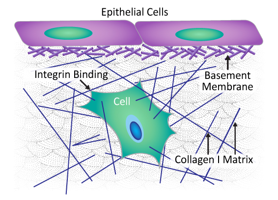
Composition and architecture of extracellular matrix (ECM). Basement membrane is a type of ECM composed of laminin and collagen IV fibers embedded within a collagen I-enriched ECM.

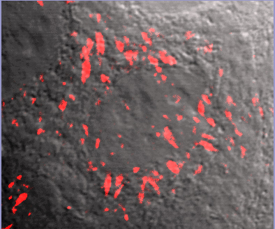
FNR01 image overlay with phase contrast background. Fluorescent fibronectin (Cat. # FNR01) treated MCF10A cells (image kindly provided by A. Varadara and M. Karthykenyan, Univ. S.Carolina, Columbia, SC).
Recently, Guzman et al. characterized a novel 3D in vitro model of multicellular cancer cell invasion that offers adjustable physiological and biochemical parameters. Shortcomings of current models are poor microscopic imaging dynamics and inability to study transmigration of cancer cells across the basement membrane (BM) and then invasion of an extracellular matrix (ECM), as happens in vivo. The BM is a type of ECM that surrounds a tumor, formed in a multi-step process initiated by cancer cells binding laminin at the cell surface to form a scaffold upon which type IV collagen polymers form. This new model offers improved imaging dynamics while also recapitulating in vivo tumor cytoarchitecture with an intact, degradable, cell-assembled, sheet-like BM layer embedded in a collagen I-enriched ECM. Cytoskeleton’s HiLyte488-conjugated laminin (Cat. # LMN02) was used to confirm that in vitro BM layer formation paralleled the in vivo process; that is, the laminin scaffold was dispersed across the surface of the multicellular tumor spheroids in a thin, patchy layer, serving as the foundation of the BM layer. This innovative 3D cancer cell invasion model offers researchers the ability to both tease apart molecular mechanisms underlying invasion and test new therapeutics in a physiologically- and biochemically-relevant setting.
