October 2020 Newsletter: Mitotic Spindle - New Tools for Visualization
- By Cytoskeleton Inc. - Live Cell News
- Oct 5, 2020
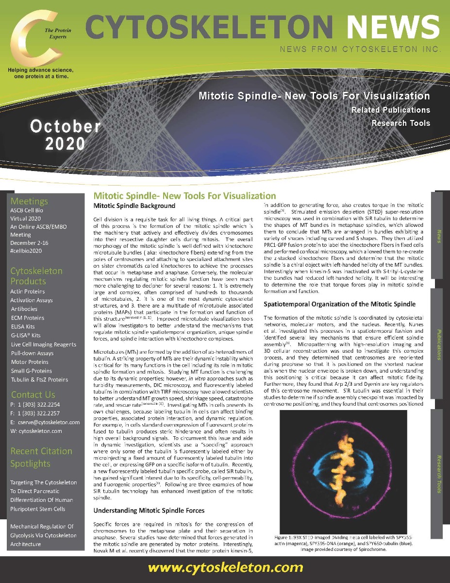
, SPY595-DNA (orange), and SPY650-tubulin (blue). Image provided courtesy of Spirochrome")
Cell division is a requisite task for all living things. A critical part of this process is the formation of the mitotic spindle which is the machinery that actively and effectively divides chromosomes into their respective daughter cells during mitosis. The overall morphology of the mitotic spindle is well-defined with kinetochore microtubule bundles ( aka: kinectochore fibers) extending from the poles of centrosomes and attaching to specialized attachment sites on sister chromatids called kinetochores to achieve the processes that occur in metaphase and anaphase. Conversely, the molecular mechanisms regulating mitotic spindle function have been much more challenging to decipher for several reasons: 1. it is extremely large and complex, often comprised of hundreds to thousands of microtubules, 2. it is one of the most dynamic cytoskeletal structures, and 3. there are a multitude of microtubule associated proteins (MAPs) that participate in the formation and function of this structure(reviewed in (1, 2)). Improved microtubule visualization tools will allow investigators to better understand the mechanisms that regulate mitotic spindle spatiotemporal organization, unique spindle forces, and spindle interaction with kinectochore complexes.
Also included in this newsletter:
- Tubulin and Actin Live Cell Reagents, G-LISA Activation Assay Kits, Tubulin Kits, Actin Biochem Kits
- Related Publications