Citation Spotlight:Transient Calcium Waves, Actin Dynamics, and Wound Healing
- By Cytoskeleton Inc. - Actin News
- Dec 3, 2019

Recently, Lee et al. used a variety of in vitro and ex vivo experimental techniques, as well as custom MATLAB analyses, to investigate transient calcium waves as a means of cell-to-cell communication to coordinate the collective migration of human corneal limbal epithelial (HCLE) cells in wound healing. Following injury, cells release the nucleotide ATP which binds P2X7 and P2Y2 purinergic receptors, triggering release of calcium and activation of associated mechanotransduction signaling pathways. The authors report that the injury-induced, sustained calcium mobilizations are mediated by pannexin channels, allowing epithelial cells to communicate and coordinate changes in the cells’ actin-based morphology to enable migration to the injury site. Calcium mobilizations are correlated with these morphological changes and subsequent increased motility, both of which require dynamic re-organization of the actin cytoskeleton. Cytoskeleton’s SiR-actin live cell imaging probe (Cat.# CY-SC001) was essential in this study as it provided a sensitive and specific method to perform short and long-term live cell confocal imaging of morphological changes in the actin cytoskeleton of HCLE cells during pharmacological manipulation of the pannexin-supported calcium mobilizations to study how different populations of HCLE cells respond to injury and participate in wound healing.
Products used in this citation:
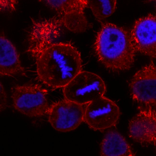
HeLa cells expressing H2B-mCherry (Blue) stained with SiR-actin (red). Image taken by confocal microscopy. Courtesy of Daniel Gerlich and Claudia Blaukopf, Institute of Molecular Biotechnology, Vienna.