The Cytoskeleton’s Role In Autism Spectrum Disorder

Introduction
 
Autism, clinically known as autism spectrum disorder (ASD), is a heritable and heterogeneous neurodevelopmental disorder with core symptoms that include difficulties with social interaction and communication, as well as repetitive behavior(1).  ASD is thought to be quite common with a prevalence approaching 1% in the worldwide population; additionally, due to its broad disease spectrum and sometimes ambiguous symptoms, it is likely that many people go undiagnosed.  Defining the underlying genetic and molecular mechanisms responsible for ASD has been elusive, and currently no single gene mutation accounts for more than 1% of ASD cases.  In support of this, a recent study, which investigated more than 35,000 human samples by whole exosome sequencing, identified 102 high-risk genes of which 53 were strongly associated with ASD(2).  These investigators aligned these high-risk ASD associated genes into 4 functional groups: 1. Gene expression and regulation, 2. Neuronal communication, 3. Cytoskeleton, and 4. Other.  The cytoskeleton has been shown to play a critical role in learning and development as well as neuronal development; thus, it is not surprising that dysfunction in cytoskeletal genes may play a fundamental role in ASD.  Below we summarize findings of three distinct ASD high-risk genes and the recent studies that highlight how they dysregulate the cytoskeleton and contribute to ASD progression.
 
Cytoskeletal Proteins, high-risk ASD genes
 
In the recent whole exosome study highlighted above, six cytoskeleton genes (GFAP, CORO1A, PYT7, DYS2A, MAP1A, and SPASTN) were identified as predominant ASD high-risk targets, but surprisingly very few studies have been performed to understand the mechanistic role for these proteins in ASD.  A recent study characterized the C58/J inbred mouse strain as a potential ASD mouse model(3).  This mouse line displays low sociability, impaired communication, and stereotyped behavior, which aligned well with the core features of ASD.  They found distinct changes in dendritic spine density and morphology and using in silico studies identified single nucleotide polymorphisms in MAP1A and other genes.  As the association between these cytoskeletal related genes and their links to ASD have only recently been identified, it will be interesting to learn about the pathways, processes, and mechanisms they modulate to induce ASD.

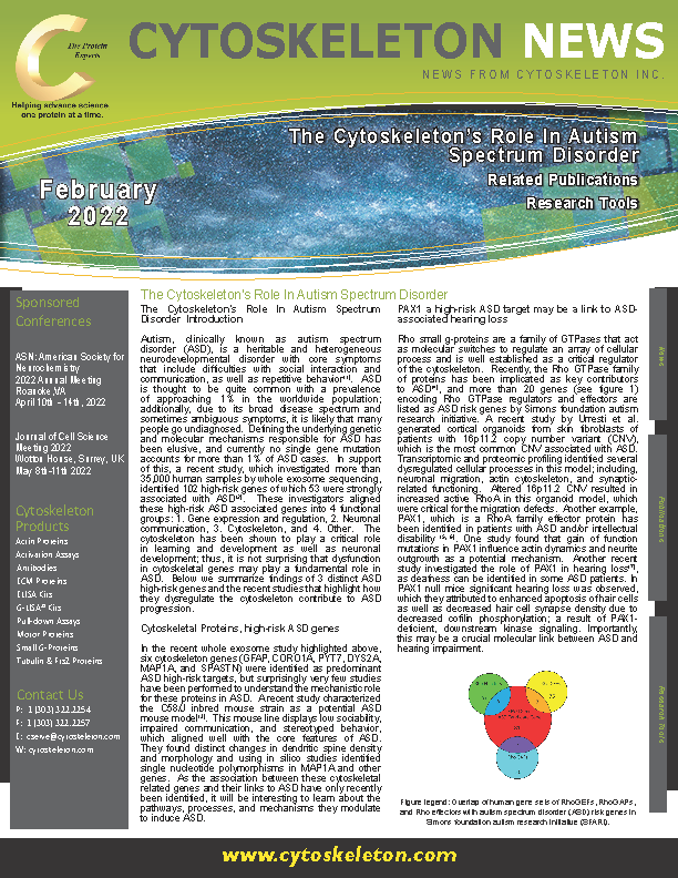
Above: Overlap of human gene sets of RhoGEFs, RhoGAPs, and Rho effectors with autism spectrum disorder (ASD) risk genes in Simons foundation autism research initiative (SFARI).

PAX1 a high-risk ASD target may be a link to ASD-associated hearing loss
 
Rho small g-proteins are a family of GTPases that act as molecular switches to regulate an array of cellular process and is well established as a critical regulator of the cytoskeleton.  Recently, the Rho GTPase family of proteins has been implicated as key contributors to ASD(4), and more than 20 genes (see figure 1) encoding Rho GTPase regulators and effectors are listed as ASD risk genes by Simons foundation autism research initiative. A recent study by Urresti et al. generated cortical organoids from skin fibroblasts of patients with 16p11.2 copy number variant (CNV), which is the most common CNV associated with ASD.  Transcriptomic and proteomic profiling identified several dysregulated cellular processes in this model; including, neuronal migration, actin cytoskeleton, and synaptic-related functioning.  Altered 16p11.2 CNV resulted in increased active RhoA in this organoid model, which were critical for the migration defects.  Another example, PAX1, which is a RhoA family effector protein has been identified in patients with ASD and/or intellectual disability (5, 6). One study found that gain of function mutations in PAX1 influence actin dynamics and neurite outgrowth as a potential mechanism.  Another recent study investigated the role of PAX1 in hearing loss (7), as deafness can be identified in some ASD patients. In PAX1 null mice significant hearing loss was observed, which they attributed to enhanced apoptosis of hair cells as well as decreased hair cell synapse density due to decreased cofilin phosphorylation; a result of PAX1-deficient, downstream kinase signaling. Importantly, this may be a crucial molecular link between ASD and hearing impairment. 
Cullin 3 modulates downstream cytoskeletal targets in ASD
 
Several other prominent genetic screens have been performed to identify causal or high-risk genes associated with ASD that has produced hundreds of additional candidates involved in processes like transcription, chromatin remodeling, and synapse regulation(8, 9).  One protein of interest Cullin 3 (Cul3) was identified in multiple studies (10, 11) but interestingly is an E3 ubiquitin ligase and does not specifically fall within the aforementioned cellular processes. So how does Cul3 regulate autism?  A recent study by Morandell et al. determined that Cul3 is critical for early neuronal development and plays a critical role in neuronal migration (12).  Specifically, it regulates levels of Plastin 3 to control the speed of neuronal migration. In their study, Cul3 deficiency led to disorganized actin architecture at the cell front, and this effect was mimicked by Plastin 3 overexpression. Another group also studying Cul3 haploinsufficiency determined using transcriptomic and proteomic profiling that the cytoskeletal defects was a key driver in Cul3 functional impact in this ASD model (13).  In their model they found reduced actin puncta in Cul3 deficient mouse neuronal cells.  Interestingly, they observed higher levels in total and active RhoA, and inhibition of RhoA was sufficient to reverse the adverse phenotypes. These studies highlight the importance of Cul3 in early neuronal development and identify multiple downstream targets that converge on the actin cytoskeleton, identifying a critical axis that may be disrupted in ASD.
 
Summary
 
Autism is clearly a complex disease that requires the dysregulation and modulation of a multitude of genes, signaling pathways, and cellular processes that lead to impaired social behavior and communication.  The highlighted studies describe a very narrow focus of recent work seeking to define the molecular underpinnings that may contribute to ASD progression with a particular focus on the role of the cytoskeleton.  Clearly the normal function of the cytoskeleton is  disrupted in ASD; however, there is still much to learn about the role of the cytoskeleton in ASD; for example, whether or not disruption of specific cytoskeletal targets alone sufficient to drive ASD and are they suitable targets for intervention. 

References:

1.Lord C, Brugha TS, Charman T, Cusack J, Dumas G, Frazier T, et al. Autism spectrum disorder. Nat Rev Dis Primers. 2020;6(1):5.
2.Satterstrom FK, Kosmicki JA, Wang J, Breen MS, De Rubeis S, An JY, et al. Large-Scale Exome Sequencing Study Implicates Both Developmental and Functional Changes in the Neurobiology of Autism. Cell. 2020;180(3):568-84 e23.
3.Baron-Mendoza I, Maqueda-Martinez E, Martinez-Marcial M, De la Fuente-Granada M, Gomez-Chavarin M, Gonzalez-Arenas A. Changes in the Number and Morphology of Dendritic Spines in the Hippocampus and Prefrontal Cortex of the C58/J Mouse Model of Autism. Front Cell Neurosci. 2021;15:726501.
4.Guo D, Yang X, Shi L. Rho GTPase Regulators and Effectors in Autism Spectrum Disorders: Animal Models and Insights for Therapeutics. Cells. 2020;9(4).
5.Horn S, Au M, Basel-Salmon L, Bayrak-Toydemir P, Chapin A, Cohen L, et al. De novo variants in PAK1 lead to intellectual disability with macrocephaly and seizures.Brain.2019;142(11):3351-9.
6.Kernohan KD, McBride A, Hartley T, Rojas SK, Care4Rare Canada C, Dyment DA, et al. p21 protein-activated kinase 1 is associated with severe regressive autism, and epilepsy. Clin Genet. 2019;96(5):449-55.
7.Cheng C, Hou Y, Zhang Z, Wang Y, Lu L, Zhang L, et al. Disruption of the autism-related gene Pak1 causes stereocilia disorganization, hair cell loss, and deafness in mice. J Genet Genomics. 2021;48(4):324-32.
8.De Rubeis S, He X, Goldberg AP, Poultney CS, Samocha K, Cicek AE, et al. Synaptic, transcriptional and chromatin genes disrupted in autism. Nature. 2014;515(7526):209-15.
9.Iossifov I, O'Roak BJ, Sanders SJ, Ronemus M, Krumm N, Levy D, et al. The contribution of de novo coding mutations to autism spectrum disorder. Nature. 2014;515(7526):216-21.
10.Nakashima M, Kato M, Matsukura M, Kira R, Ngu LH, Lichtenbelt KD, et al. De novo variants in CUL3 are associated with global developmental delays with or without infantile spasms. J Hum Genet. 2020;65(9):727-34.
11.Sanders SJ, He X, Willsey AJ, Ercan-Sencicek AG, Samocha KE, Cicek AE, et al. Insights into Autism Spectrum Disorder Genomic Architecture and Biology from 71 Risk Loci. Neuron. 2015;87(6):1215-33.
12.Morandell J, Schwarz LA, Basilico B, Tasciyan S, Dimchev G, Nicolas A, et al. Cul3 regulates cytoskeleton protein homeostasis and cell migration during a critical window of brain development. Nat Commun. 2021;12(1):3058.
13.Amar M, Pramod AB, Yu NK, Herrera VM, Qiu LR, Moran-Losada P, et al. Autism-linked Cullin3 germline haploinsufficiency impacts cytoskeletal dynamics and cortical neurogenesis through RhoA signaling. Mol Psychiatry. 2021;26(7):3586-613.