H001 has been discontinued, this page remains for reference and information.
* Limited stock available. If stock is not available, Cytoskeleton will produce a new batch upon request. Minimum order will apply. Inquire for more information.
Product Uses Include
- Screening for anti-tumor drugs
- Assessing the effects of modulators of microtubule dynamics on tubulin isolated from actively growing cells
- A new substrate for motor proteins
Material
HeLa Cell tubulin is isolated from the human cervical cancer derived HeLa S3 cell line, a model system to study many aspects of tumor cell growth. Hela Cell tubulin may be used in all situations where previously bovine brain tubulin has been employed, for example drug screening, motility assays and biochemical studies including microtubule dynamics. The advantage of using this novel tubulin is that it is derived from an actively dividing cell line which often, in contrast to brain derived tubulins, more accurately portrays the situation that many researchers are trying to reconstruct in vitro.
The specificity of ligands for for a particular tubulin variant can be determined by performing comparative studies with both cancer cell and neuronal tubulins. Cytoskeleton, Inc. has advanced this concept by developing the Tubulin Ligand Index (TLI) system (patent pending). In this system, IC50 values for inhibitory compounds or EC50 values for stabilizing compounds are determined in in polymerization assays using cancer cell and neuronal tubulins. The IC50 or EC50 values for each tubulin variant are analyzed as a ratio (neuronal/cancer cell) and allow for determinations of the relative specificity for each tested compound. TLI values greater than 1.0 indicate that the particular compound is more active on cancer cell tubulin. Conversely, TLI values less than 1.0 suggest that a compound is more specific for neuronal tubulin. Table 1 summarizes data from a study comparing the specificity of several tubulin ligands using the TLI system.
HeLa cell tubulin is also available as a biotinylated conjugate for scintillation proximity assay (SPA) applications (Cat. # H003). For another human cancer cell derived tubulin, see our MCF-7 tubulin (Cat. # H005) Purity H001 contains >90% pure HeLa cell tubulin. Purity is determined by scanning densitometry of proteins on SDS-PAGE gels.
| Table 1. Tubulin ligand index values from studies with cancer cell and neuronal tubulins | ||||||
EC50/IC50 Value (µM) | TLI Value | |||||
Ligand | Neuronal | MCF-7 | HeLa | MCF-7 | HeLa | |
Paclitaxel Docetaxel 10-Deacetyl Taxol | 0.48 0.47 3.71 | 0.51 0.34 4.20 | 1.04 0.41 30.00 | 0.94 1.38 0.88 | 0.46 1.15 0.12 | |
Vinblastine Vincristine | 1.10 1.58 | 1.21 n.d. | 2.83 2.25 | 0.91 n.a. | 0.39 0.70 | |
Colchicine Nocodazole Mebendazole MF708 | 4.10 3.40 3.98 3.54 | 4.60 3.20 14.80 n.d. | 3.10 3.20 25.00 1.91 | 0.89 1.06 0.27 n.a. | 1.32 1.06 0.16 1.85 | |
Cytoskeleton, Inc's HeLa cell tubulin protein is provided as a lyophilized powder and when reconstituted, it is in 80 mM PIPES, 1 mM MgCl2, 1 mM EGTA and 1 mM GTP (G-PEM), pH 6.9.
HeLa cell tubulin is also available as a biotinylated conjugate for scintillation proximity assay (SPA) applications (Cat. # H003). For another human cancer cell derived tubulin, see our MCF-7 tubulin (Cat. # H005)
Purity
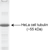
H001 contains >90% pure HeLa cell tubulin. Purity is determined by scanning densitometry of proteins on SDS-PAGE gels.

Figure 1: An H001 sample (20 µg) was run on a 10% SDS-PAGE gel and stained with Coomassie Blue stain
Biological Activity
One unit of tubulin is defined as 24 µg of purified protein (as determined by the Precision Red Advanced Protein Assay Reagent, Cat. # ADV02). HeLa cell tubulin will polymerize efficiently at a concentration of 2.0 mg/ml in the presence of 20% (v/v) glycerol. The HeLa tubulin polymerization assay has been miniaturized to 12 µl (1 unit/reaction) in order to make anti-cancer drug development feasible. Visit the page on CytoDYNAMIX screens for more information. HeLa cell tubulin is also available as a biotinylated conjugate for Scintillation Proximity Assay applications (Cat. # H003). Tubulin from a breast cancer cell line (MCF-7) is also available (Cat. # H005).
For product Datasheets and MSDSs please click on the PDF links below. For additional information, click on the FAQs tab above or contact our Technical Support department at tservice@cytoskeleton.com
Question 1: Can I study the in vitro polymerization of HeLa cell tubulin?
Answer 1: Yes, HeLa cell tubulin (Cat. # H001) can be used in polymerization assays using either the turbidometric tubulin polymerization kit (Cat. # BK006P) or the fluorescent tubulin polymerization kit (Cat. # BK011P). The HeLa cell tubulin is substituted for the porcine tubulin included in the kit. HeLa cell tubulin polymerization into microtubules can be detected by measuring the optical density (OD) at 340 nm or by measuring the change in fluorescence intensity in the presence of DAPI at excitation and emission wavelengths of 360 nm and 405-450 nm, respectively. The fluorescence technique is recommended over the absorbance technique due to the expensive nature of the protein and the small volume possible with a fluorescence format (i.e. 10 µl, see datasheet for more information).
Question 2: What is the advantage of using HeLa cell tubulin vs mammalian brain tubulin?
Answer 2: Mammalian brain tubulin (Cat. # T240) is useful for an initial or primary screening of anti-cancer compounds and for preliminary determinations of a compound’s IC50 values. However, for more detailed, specific information on how the anti-cancer compounds will actually affect cancer cell tubulin, we recommend using either the HeLa cell tubulin (Cat. # H001) or MCF-7 tubulin (Cat. # H005). It is well-known that a drug’s efficacy and binding dynamics can vary between neuronal tubulin and cancer cell tubulin. In addition, many of the side effects associated with anti-cancer treatments are attributed to the compounds’ effects on neuronal tubulin. Therefore, to insure the most specific and selective drug-target interactions, we recommend secondary screens with cancer cell tubulins.
If you have any questions concerning this product, please contact our Technical Service department at tservice@cytoskeleton.com


