Live Cell Imaging Tools
Cytoskeleton offers several reagents for live-cell research including fluorescent proteins, cell permeable protein activators and inhibitors, as well as our recent addition of live cell imaging probes.
Learn More About Live Cell Imaging
Our most recent Live Cell writings can be found below. However Cytoskeleton has been writing about Live Cell Imaging for years, and that library of information can be found here
Malaria Influences The Cytoskeleton During It’s Lifecycle
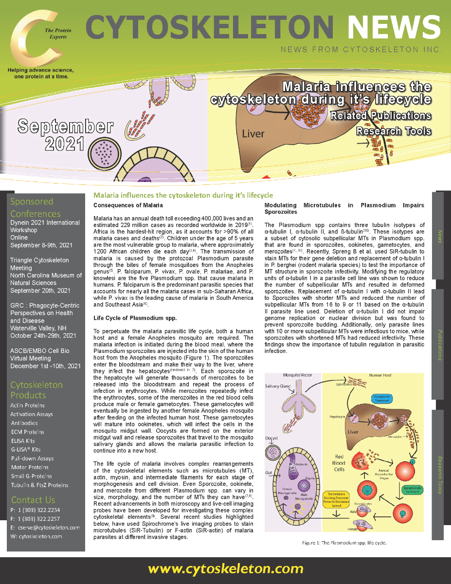
Malaria has an annual death toll exceeding 400,000 lives and an estimated 229 million cases as recorded worldwide in 2019(1). Africa is the hardest-hit region, as it accounts for >90% of all malaria cases and deaths(2). Children under the age of 5 years are the most vulnerable group to malaria, where approximately 1200 African children die each day(3,4). The transmission of malaria is caused by the protozoal Plasmodium parasite through the bites of female mosquitoes from the Anopheles genus(5). P. falciparum, P. vivax, P. ovale, P. malariae, and P. knowlesi are the five Plasmodium spp. that cause malaria in humans. P. falciparum is the predominant parasitic species that accounts for nearly all the malaria cases in sub-Saharan Africa, while P. vivax is the leading cause of malaria in South America and Southeast Asia(6).
Investigating Exosomes - Tools And Implications
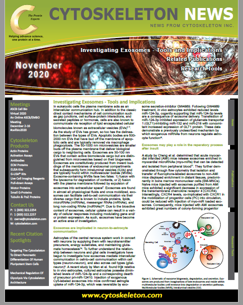
In eukaryotic cells the plasma membrane acts as an intercellular communication hub. In addition to the classic direct contact mechanisms of cell communication such as gap junctions, cell surface-protein interactions, and secreted peptides or hormones, cells are also known to communicate via reception of lipid encapsulated cellular biomolecules known as extracellular vesicles (EVs)1–3. As the study of EVs has grown, so too has the delineation between the types of EVs. Apoptotic bodies are 500-2000 nm EVs that have bud off the membrane of apoptotic cells and are typically removed via macrophage phagocytosis. The 50-1000 nm microvesicles are smaller buds off the plasma membrane that deliver biological cargo to neighboring cells. Exosomes are 30-100 nm EVs that contain active biomolecule cargo but are distinguished from microvesicles based on their biogenesis.
Live Cell Imaging Tools In Action - Citation Spotlights
Plexin-B2 Orchestrates Collective Stem Cell Dynamics Via Actomyosin Contractility, Cytoskeletal Tension And Adhesion
Pro-Inflammatory Stressors Alter The Vesiculome Of MIN6 Pancreatic Beta Cells
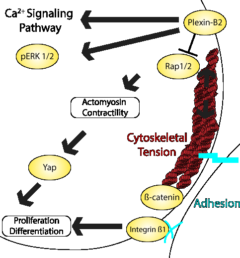
Multicellular organization requires rapid changes in biomechanical dynamics and establishment of a collective actomyosin network in order to maintain cell morphology. How human embryonic stem cells (hESCs) orchestrate cytoskeletal tension across cells and organize adhesion complexes to maintain cytoarchitectural integrity is incompletely understood. Recent work by the Friedel lab identified a key role for Plexin-B2 in the control of actomyosin contractility and adhesion properties in hESCs. Plexin-B2 is an axon guidance receptor that has been shown to mediate cellular interactions in a variety of tissue contexts. The group utilized Plexin-B2 CRISPR KO hESCS (B2-KO) as well as lentiviral Plexin-B2 overexpressing hESCs [B2-OE] to show that dysregulation of Plexin-B2 severely alters hESC colony expansion and geometry. Utilizing RNA-Seq approaches they identified pathways related to muscle contraction, ECM organization, integrin signaling, and elastic fiber formation to be particularly augmented. A deeper investigation into the morphological changes revealed significant alterations in both F-actin networks and phosphorylated myosin light chain 2 localization. ...
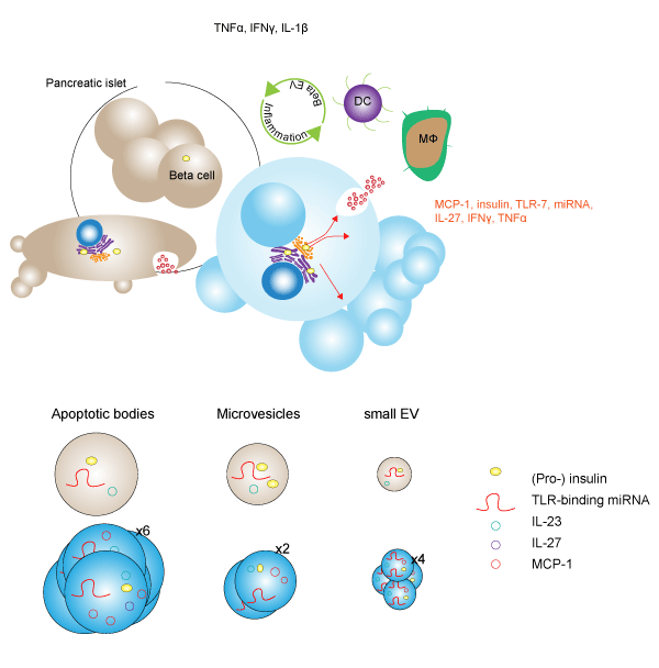
The development of autoimmune type 1 diabetes is linked to chronic inflammation of pancreatic beta islet cells. While smaller extracellular vesicles (EVs), such as exosomes, have been implicated as key contributors to the development of pathogenic-inflammation in beta cells, the contribution of larger EVs such as apoptotic bodies (ABs), microvesicles (MVs), and small EVs (sEVs) is unknown. In a recent study, Giri et al. treated murine MIN6 beta cells with pro-inflammatory stressors such as incubation in 1% O2 (HX), ultraviolet irradiation (UV), and exposure to pro-inflammatory cytokines (CK), and characterized changes in the vesicular secretome, or vesiculome. Following treatment, investigators used size exclusion-based methods to isolate putative EVs. Lipid bilayer membrane analysis was performed with MemGlow in conjunction with cryo-electron microscopy to confirm EV capture. Examination of EVs with Tunable Resistive Pulse Sensing (TRPS) revealed that while stress-treatments of MIN6-derived EVs did not significantly change the size of individual vesicles, the number of EVs significantly increased ABs (5.5-fold), MVs (2.1-fold), and sEVs (4.5-fold) resulting in an overall increased vesiculome volume in a stress-dependent manner...
Please visit our product pages for product specific citations
SiR-DNA Kit - MemGlow™ 488: Fluorogenic Membrane Probe - Flourescent HiLyte Tubulin Protein
Cytoskeleton's line of Live Cell Imaging Tools
Live Cell Imaging Tools Brochure
Browe Cytoskeleton Inc's selection of tools by clicking the brochure above, or the button below which will take you to a category page which you can navigate to see our full selection of Live Cell imaging Tools.
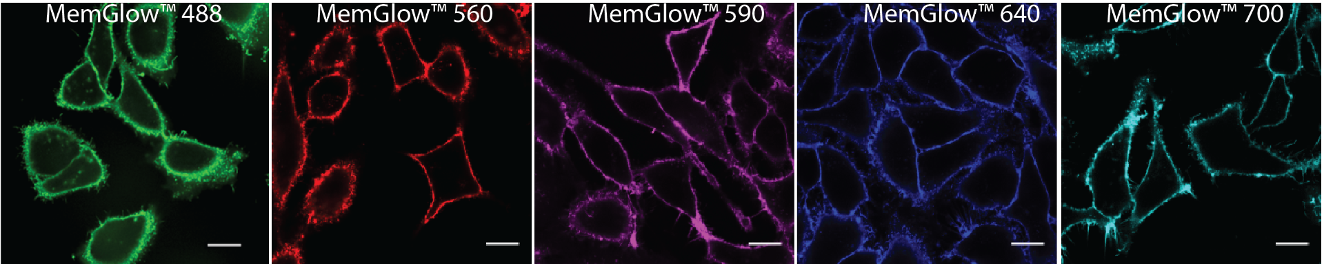
Originally developed as the MemBright™ probes, MemGlow™ probes provide high photostability, exhibit excellent fluorescent quantum yields, and some of them are super-resolution compatible. Structurally, MemGlow™ probes are composed of cyanine or BODIPY dyes bearing zwitterionic amphiphilic anchors for enhanced retention in the plasma membrane.
Have a technical question about our tools? Send us an email at tservice@cytoskeleton.com and our experts will anwser any question you may have!