Figure 2. Absorbance and excitation spectra of MemGlow NR12S diluted in DMSO to 4.6 µM. Absorbance peak detected at 550 nm with peak emission at 633 nm.
MemGlow NR12S is a solvatochromic photostable plasma membrane-targeting dye. Upon binding with a plasma membrane in a predominant liquid ordered (LO) phase NR12S exhibits a 45-50 nm wavelength shift relative to liquid disordered (LD) phase enabling investigators to examine the nanoscale distribution of local chemical polarity in plasma membranes. NR12S can also be used to label the plasma membrane for conventional fluorescence microscopy imaging.
NR12S Membrane Polarity Probe Product Functions (MG08)
-
MemGlow NR12S is a solvatochromic photostable plasma membrane-targeting dye
-
Examine the nanoscale distribution of local chemical polarity in plasma membranes
-
NR12S can also be used to label the plasma membrane for conventional fluorescence microscopy imaging.
For more detailed information on using this product see the About Tab
** 4 nmol of MemGlow will produce a 20µM stock solution when reconstituted with 200 µl of anhydrous DMSO
.")
Figure 3. Widefield fluorescent imaging of live HeLa S1 cells labeled with 10 nM NR12S. HeLa S1 cells were imaged with a TRITC filter set, neutral density filter, a digital CCD camera, and 100x oil objective (false colored red).

Figure 1. Diagram of NR12S’s lipid order-dependent emission.
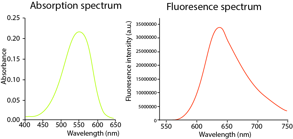

Figure 3. Fluorescence spectra of NR12S in Lo (Cholesterol) and Ld (DOPC) phases.
Material
The absorption max of NR12S is 554 nm, with an emission spectra of 635 nm, an extinction coefficient of 45,000 M-1.cm-1 , and can be visualized using a Cy 3.5 filter set or other suitable filter sets. NR12S is supplied as a lyophilized pellet. NR12S is a lipid binding dye and appropriate PPE should be worn at all times. Dispose of NR12S according to local regulations and policies.
Storage and Reconstitution
The lyophilized product is stable at 4°C (<10% humidity) for 6 months and should be protected from light. To reconstitute, briefly centrifuge to collect the product at the bottom of the tube. NR12S should be reconstituted with 200 µl of anhydrous DMSO to create a 20 µM stock solution for cell imaging. After reconstitution the solution should be stored at -20°C where it is stable for 3 months. Once reconstituted, allow product to warm to room temperature before opening tube.
Important Technical Notes
- NR12S was purpose-engineered for super resolution techniques. It is not intended for use in conventional microscopy; however, it can be used with good results.
- Diluted solutions of NR12S in aqueous media should be used as soon as possible as it may precipitate.
- Serum and proteins will reduce NR12S staining efficiency. When possible NR12S staining should take place in the absence of serum. Optimally, the imaging cell media is serum-free media, reduced serum media, or PBS.
- NR12S is generally non-toxic to live cells but morphological changes 12 hours after no-wash applications can cause cell shrinkage. For repeated imaging, follow cell labeling with a wash using cell media.
- When co-labeling with antibodies that require permeabilization limit the concentration of Triton-X to 0.1%.
- NR12S will work with a range of concentrations. Investigators should empirically determine the best concentration for their application. An initial concentration range of 50-200 nM is recommended.
- NR12A is a next generation version of the NR12S probe and has more than 2-fold higher affinity to the plasma membrane.
Table 1. Recommended initial concentrations. Optimal conditions for efficient labeling should be determined for each cell line and application.
Reagents
- NR12S (Cat. # MG08).
- Semi-confluent HEK293 cells grown in a chamber slide.
- Imaging medias: PBS, serum-free media or reduced serum media.
Equipment
- Fluorescent microscope with a Cy 3.5 excitation filter at 550 +/-20 nm and emission filter at 633 +/-20 nm for NR12S.
- Digital camera.
Method
- Cells should be seeded onto imaging-appropriate glass or plastics and grown according to cell line requirements to semi-confluency.
- Remove any cell culture media from your cells and replace with the media used for imaging (e.g., serum-free media). Do not allow the cells to dry.
- Prepare the probe solution by diluting 5 µl of 20 µM MemGlow NR12S stock in 1 mL imaging media to create a 100 nM working solution or and mix thoroughly. Work quickly as the probes will begin to aggregate over time reducing labeling efficiency.
- Replace the cell media with diluted probe solution ensuring cells are submerged. Incubate cells in MemGlow™ solution for 10 minutes at room temperature.
- For SR-Paint applications the cells are ready to image. For general microscopy applications no washing step is required prior to imaging, but can be performed if desired with imaging media.
Product Citations
- Kucherak, O. A. et al. Switchable nile red-based probe for cholesterol and lipid order at the outer leaflet of biomembranes. J. Am. Chem. Soc. 132, 4907–4916 (2010).
- Danylchuk, D. I., Moon, S., Xu, K. & Klymchenko, A. S. Switchable Solvatochromic Probes for Live-Cell Super-resolution Imaging of Plasma Membrane Organization. Angew. Chemie - Int. Ed. 58, 14920–14924 (2019).
MEMBRIGHT™ is a trademark of CNRS/UNISTRA of France.