PKmito RED is a bright, non-phototoxic & non-toxic mitochondrial probe based on the PKmito™ dyes developed by by Yang et al in the lab of Zhixing Chen1). PKmito RED labels mitochondria in live cells with very high specificity. The unique and unmatched feature of PKmito RED is its extremely low phototoxicity, due to the presence of the intramolecular triplet quencher cyclooctatetraene (COT) group. It allows to perform long term imaging of mitochondria without damaging them. PKmito RED accumulates in the mitochondrial inner membrane (IM) and can used to image mitochonridal cristae by STED or SIM superresolution microscopy. PKmito RED does not require any genetic manipulation, transfection or overexpression of fluorescent proteins. PKmito RED enables multicolor imaging with SPY505, SPY595, SPY650, SPY700, SiR or GFP. PKmito RED can be imaged with a standard TMR or Cy3 filterset. It can be used for widefield, confocal, SIM or STED imaging in living cells and tissue. Contains 1 vial of PKmito RED (lyophilized).
Absorbance maximum λabs | 549 nm |
Fluorescence maximum λfl | 569 nm |
Works on fixed cells? | No |
Probe quantity | 100 stainings* |
Fluorescence lifetime | 0.8 ns |
STED depletion wavelength | 660 nm |
Shipping | room temperature |
Storage | -20°C |

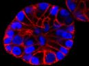
Cells Stained with PKmito RED

PKmito RED
For product Datasheets and MSDSs please click on the PDF links below.
Spirochrome Technical Tips and Ex/Em spectra in graphical form (PDF)
If you have any questions concerning this product, please contact our Technical Service department at tservice@cytoskeleton.com
Q1. What is STED microscopy and how does it work?
A1. STED microscopy stands for Stimulated Emission Depletion microscopy. It is one type of super resolution microscopy which allows the capture of images with a higher resolution than conventional light microscopy which is constrained by diffraction of light. STED uses 2 laser pulses, one is the excitation pulse which excites the fluorophore, causing it to fluoresce. The second pulse, referred to as the STED pulse, de-excites the fluorophore via stimulated emission in an area surrounding a central focal spot that is not de-excited and thus continues to fluoresce. This is accomplished by focusing the STED pulse into a ring shape, a so-called donut, where the center focal spot is devoid of the STED laser pulse, conferring high resolution to the fluorescent area (Fig. 1; see Ref. 1 for more details on STED microscopy).
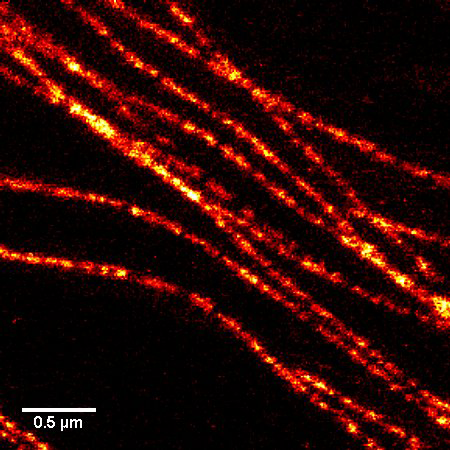
Figure 1. STED microscopic image of microtubules labeled with SiR-tubulin in human primary dermal fibroblasts.
Q2: Are the SPY™ probes stable at room temperature?
A2: Yes, the probes are stable at room temperature for a few days. However, it strongly depends on the probe and the solvent. Thus, it is recommended to store all of the probes or solutions at –20°C.
References
1. Liu Tianyan et al. “Multi-color live-cell STED nanoscopy of mitochondria with a gentle inner membrane stain” PNAS (2022):119 (52).
2. Yang, Zhongtian, et al. “Cyclooctatetraene-conjugated cyanine mitochondrial probes minimize phototoxicity in fluorescence and nanoscopic imaging.” Chemical science 11.32 (2020): 8506-8516.