SPYRHO 555 is a fluorogenic dye ligand for the RNA aptamer RhoBAST. SPYRHO 555 was developed by the lab of Murat Sunbul in Heidelberg University. SPYRHO 555 is cell permeable, highly fluorogenic and photostable. With its high affinity and fast on/off binding kinetics, it allows overcoming the main issues of live cell RNA imaging. SPYRHO 555 is highly suited for widefield, confocal, STED or single molecule localization microscopy. SPYRHO 555 can be imaged with standard TMR or Cy3 filtersets. It can be used in living or fixed cells. Contains 1 vial of SPYRHO 555 (lyophilized).
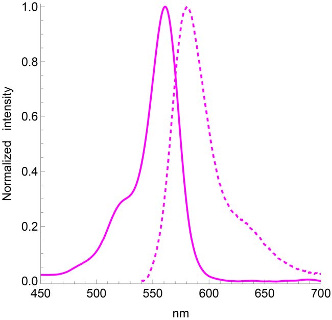
Absorbance maximum λabs | 562 nm |
Fluorescence maximum λfl | 581 nm |
Works on fixed cells? | yes |
Probe quantity | 100 stainings* |
ΦF (RNA bound) | 0.95 |
Kd to RhoBAST aptamer | 34nM |
STED depletion wavelength | 660 nm |
| MW | 492.5 g/mol |
Shipping | room temperature |
Storage | -20°C |
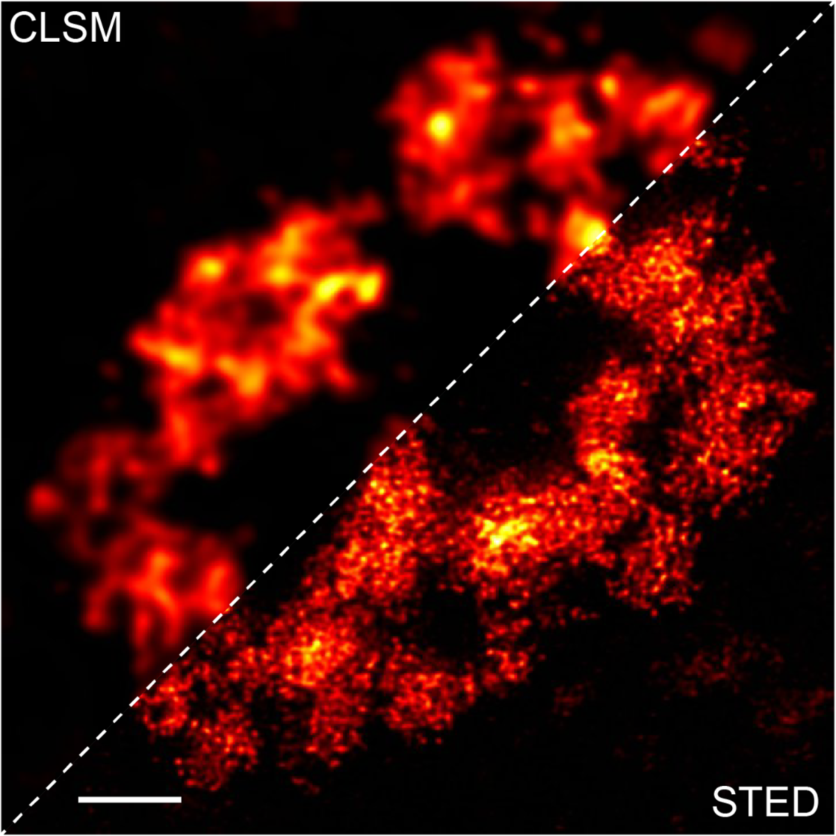
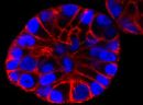
Legend: Deconvoluted CLSM and STED image of living Cos7 cells expressing CGG99-FMR1-GFP-RhoBAST16 mRNA incubated with SpyRho (100 nM) for 30 min before imaging. Scale bar, 2µm.
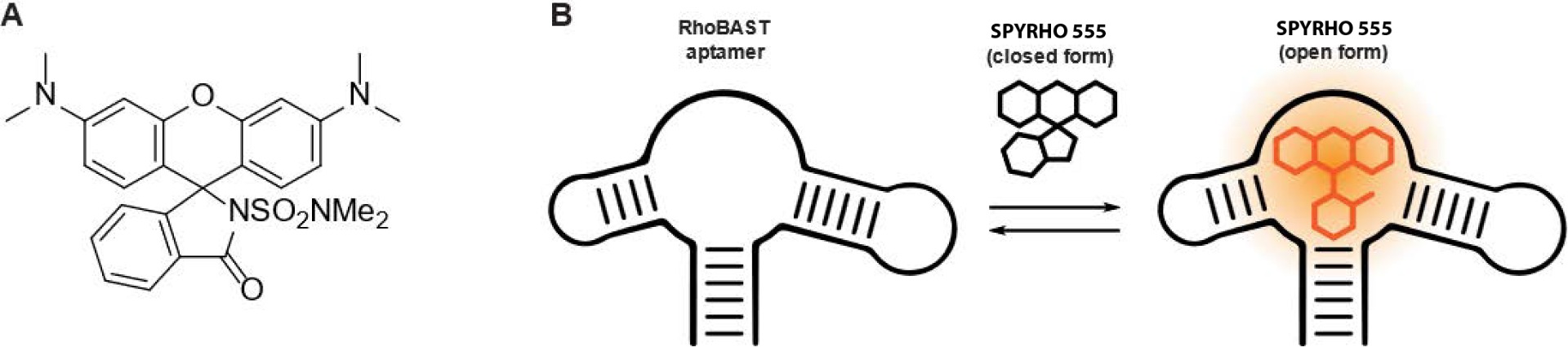
Figure 1. A Chemical structure of SPYRHO 555. B SPYRHO 555 reversibly binds to RhoBAST aptamer with fast kinetics. Unbound SPYRHO 555 exists
predominantly in the closed form and is not fluorescent. Upon binding to RhoBAST aptamer, SPYRHO 555 switches to the open form and becomes
highly fluorescent.
For product Datasheets and MSDSs please click on the PDF links below.
If you have any questions concerning this product, please contact our Technical Service department at tservice@cytoskeleton.com
Q1. What is STED microscopy and how does it work?
A1. STED microscopy stands for Stimulated Emission Depletion microscopy. It is one type of super resolution microscopy which allows the capture of images with a higher resolution than conventional light microscopy which is constrained by diffraction of light. STED uses 2 laser pulses, one is the excitation pulse which excites the fluorophore, causing it to fluoresce. The second pulse, referred to as the STED pulse, de-excites the fluorophore via stimulated emission in an area surrounding a central focal spot that is not de-excited and thus continues to fluoresce. This is accomplished by focusing the STED pulse into a ring shape, a so-called donut, where the center focal spot is devoid of the STED laser pulse, conferring high resolution to the fluorescent area (Fig. 1; see Ref. 1 for more details on STED microscopy).
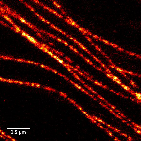
Figure 1. STED microscopic image of microtubules labeled with SiR-tubulin in human primary dermal fibroblasts.
Q2: Are the SPY™ probes stable at room temperature?
A3: Yes, the probes are stable at room temperature for a few days. However, it strongly depends on the probe and the solvent. Thus, it is recommended to store all of the probes or solutions at –20°C.
References
1. Daniel Englert et al. Fast-exchanging spirocyclic rhodamine probes for aptamer-based super-resolution RNA imaging” bioRxiv
2022.10.24.513449 https://doi.org/10.1101/2022.10.24.513449