Manipulation of Small GTPases by Pathogenic Bacteria
Small GTPases, also known as small G-proteins, are a large family of regulatory factors with roles in virtually all cellular processes. They essentially act as molecular switches, adopting either GDP-bound inactive (“off”) or GTP-bound active (“on”) states, through structural changes induced by the presence or absence of the γ-phosphate group of GTP.1 In humans, the small GTPase family contains approximately 150 members, and is further classified into five subfamilies—Ras, Rho, Arf, Rab, and Ran—based on their sequence and structure.2
The activity of these proteins is tightly regulated by a series of intricate mechanisms3 (Figure 1). Switching from the “off” to the “on” state is mediated by guanine nucleotide exchange factors (GEFs), which promote release of GDP and its replacement with GTP. Conversely, GTPase-activating proteins (GAPs) stimulate the normally slow intrinsic GTPase activity to enable rapid return to the “off” state. In this state, small GTPases are sometimes bound to guanine dissociation inhibitors (GDIs) to block interaction with GEFs and prevent activation. This elaborate control system is further enhanced by various protein–protein interactions and posttranslational modifications (PTMs),4 resulting in a highly complex regulatory network whose functioning has been the subject of intense research over the past few decades.
Given its importance, it is unsurprising to find that many pathogenic bacteria co-opt small GTPase signaling to invade and colonize host tissues. Here, we will illustrate just a few of the diverse strategies these pathogens use to subvert host cellular processes to their own advantage.
Activating Rho GTPases for Host Cell Invasion
The membrane-anchored Rho-family GTPases—including Rac1, Cdc42, and RhoA—are key regulators of actin cytoskeleton organization. These factors moderate the function of WASP and WAVE proteins to direct recruitment and activation of the actin-polymerizing Arp2/3 complex.5 Normally, WASP adopts an autoinhibitory fold where the Arp2/3-activating VCA motif is shielded by the G-protein binding domain (GBD). Association of the latter with Cdc42–GTP releases the VCA to engage Arp2/3,6 and Rac1–GTP similarly activates N-WASP and WAVE.7,8 Many pathogenic bacteria subvert Rho-family GTPase function to effect cytoskeletal remodeling and enable invasion of host cells. For example, Campylobacter jejuni (a major cause of food poisoning) hijacks an integrin-mediated signaling cascade upstream of Rac1 by binding fibronectin through two outer membrane proteins, CadF and FlpA.9 This induces downstream recruitment of host GEFs (DOCK1 and TIAM1) that activate Rac1, promoting membrane ruffling and bacterial entry. Consistent with this, knockdown of Rac1 or its GEFs largely attenuated C. jejuni invasion of human epithelial cells.
Rather than exploiting membrane receptors, some bacteria inject effector proteins directly into the host cell cytoplasm using a syringe-like type III secretion system (T3SS). This mechanism is exemplified by Salmonella species (gastroenteritis and typhoid fever), which have been extensively studied as model pathogens, and secrete a number of effectors that interfere with small GTPase function to permit bacterial internalization and intracellular survival.5 The Salmonella secretome includes SopE, a eukaryotic GEF mimic that targets Cdc42 and Rac110 to drive actin polymerization and membrane remodeling, promoting host cell invasion.
Subsequent work revealed that WAVE activation also requires Arf-family small GTPases, as well as Rac1. Through the action of a second effector, SopB, Salmonella recruits the host GEF cytohesin-2 (ARNO) to activate Arf1 and complement the function of SopE.11,12 In addition, SopE is also now known to activate the small GTPase RalA, to recruit the exocyst complex to invasion sites and further drive membrane expansion.13 Remarkably, the bacterium secretes yet another protein after entry, SptP, with GAP activity for Cdc42 and Rac1, to downregulate the membrane remodeling process and restore normal cytoskeletal morphology.14,15
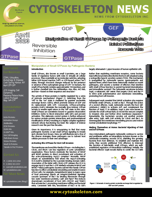

Figure. Schematic of small g-protein nucleotide exchange that is regulated by GEF and GAP proteins.
Making Themselves at Home: Bacterial Hijacking of Rab and Arf GTPases
Once internalized, pathogenic Salmonella continue to survive and proliferate within an intracellular inclusion known as the Salmonella-containing vacuole (SCV), shielding the growing bacteria from host defenses. To establish and maintain this niche, they secrete additional T3SS effectors to intercept the function of Rab-family small GTPases, which are well-established regulators of intracellular trafficking.4 Initially, the bacterial effector SopB recruits Rab5a to confer the SCV with early endosomal characteristics, including EEA1 loading.16 Since Rab5a mediates early endosomal fusion and conversion to late endosomes, the maturing SCV then acquires late endosomal markers including Rab7 and Rab9, although further T3SS effectors modify its characteristics to permit bacterial survival. For example, SifA captures additional Rab7 by binding to its effector protein, PLEKHM1, to divert phagolysosomal membranes for fusion with the SCV, thereby expanding the vacuole.17 Meanwhile, SopD2 blocks Rab7-mediated recruitment of RILP and FYCO1 to prevent trafficking of the SCV to lysosomes,16 and SifA also engages PLEKHM2 to block its interaction with Rab9. This stops delivery of mannose-6-phosphate receptors (M6PRs) to the SCV membrane, preventing import of lysosomal enzymes and protecting the resident bacteria from hydrolytic attack.18
Similarly, Chlamydia trachomatis proliferates intracellularly by manipulating multiple pathways through more than 70 T3SS-delivered proteins.19 After using IPAM/CT223 to subvert host microtubule organization and position its nascent vacuole next to the Golgi apparatus, Chlamydia secretes InaC/CT813 to recruit the Arf1 and Arf4 GTPases to the inclusion membrane. This induces stabilizing microtubule PTMs,20 seemingly “anchoring” the vacuole in place. Like other obligate intracellular bacteria, C. trachomatis has a small genome and lacks many metabolic genes, making it reliant on nutrients from the host cell. Thus, fusion of Golgi-derived vesicles, captured by the remodeled microtubule network, allows the bacteria to obtain essential lipids.21 Another chlamydial effector, CT229/CpoS, sequesters further nutrients through its ability to bind several Rab GTPases. For example, interactions with Rab4 and Rab35 enable it to intercept transferrin-containing recycling vesicles to obtain iron, an essential nutrient for productive chlamydial infection.22
Diverting Autophagy: To Exploit or Evade?
Multiple Rab GTPases are also involved in regulating autophagy,23 and different bacteria manipulate them in different ways. The intracellular pathogens Coxiella burnetii (Q fever) and Ehrlichia chaffeensis (human monocytic ehrlichiosis) are both thought to capture autophagosomes as a source of nutrients and membrane materials to support bacterial proliferation and vacuole expansion. C. burnetii achieves this by T4SS-mediated delivery of CvpF, an effector that recruits Rab26 to the inclusion.24 This is a recently characterized small GTPase that has been shown to promote autophagosome formation in several studies.25–27 In the case of E. chaffeensis, the secreted factor Etf-1 binds to both Rab5a and its effector PIK3C3. This stimulates generation of an autophagosome that subsequently associates with the bacterial inclusion, ostensibly delivering cytoplasmic nutrients to the replicative vacuole.28
Autophagy is also an important host defense mechanism against invading intracellular bacteria, but it can be subverted by certain pathogens. For example, Listeria monocytogenes (listeriosis) secretes an enzyme that ADP-ribosylates Rab5a, blocking GEF function and preventing its activation. This inhibits Rab5a-mediated fusion of phagosomes with lysosomes, enabling the bacteria to evade killing.29 Similarly, Shigella flexneri (diarrhea) disarms autophagy via the T3SS effector VirA, which functions as a GAP for Rab1 to maintain it in the “off” state and prevent autophagosome formation.30
Recent Developments & Future Prospects
Given the range of functional virulence factors released by bacterial secretion systems, targeting of these effectors—or of the T3SS itself—has been suggested as a novel therapeutic strategy.31 This might offer a potential solution to growing antibiotic resistance, as well as other adverse effects of the existing agents,32 and several new drug candidates are in preclinical development. Other work has identified a small GTPase-like domain in the Smc protein of Mycoplasma pulmonis, which structurally and functionally resembles SopE-like GEFs, and was found to activate Rac1 to promote cellular migration and proliferation.33 Long-term mycoplasma infections are currently difficult to resolve, but are associated with elevated risk of multiple cancer types,34 so the possibility that these pathogens employ bacterial-like virulence factors may offer a promising new avenue for future cancer treatment and prevention.
References
-
Toma-Fukai S, Shimizu T. Structural insights into the regulation mechanism of small GTPases by GEFs. Molecules. 2019;24(18):3308. https://doi.org/10.3390/molecules24183308.
-
Paone S, Olivieri A. Role of host small GTPases in Apicomplexan parasite infection. Microorganisms. 2022;10(7):1370. https://doi.org/10.3390/microorganisms10071370.
-
Corry J, Mott HR, Owen D. Activation of STAT transcription factors by the Rho-family GTPases. Biochem Soc Trans. 2020;48(5):2213–2227. https://doi.org/10.1042/bst20200468.
-
Cherfils J, Zeghouf M. Regulation of small GTPases by GEFs, GAPs, and GDIs. Physiol Rev. 2013;93(1):269–309. https://doi.org/10.1152/physrev.00003.2012.
-
Rottner K, Stradal TEB, Wehland J. Bacteria–host-cell interactions at the plasma membrane: Stories on actin cytoskeleton subversion. Dev Cell. 2005;9(1):3–17. https://doi.org/10.1016/j.devcel.2005.06.002.
-
Higgs HN, Pollard TD. Activation by Cdc42 and PIP2 of Wiskott–Aldrich syndrome protein (WASP) stimulates actin nucleation by Arp2/3 complex. J Cell Biol. 2001;150(6):1311–1320. https://doi.org/10.1083/jcb.150.6.1311.
-
Tomasevic N, Jia Z, Russell A, Fujii T, Hartman JJ, Clancy S, Wang M, Beraud C, Wood KW, Sakowicz R. Differential regulation of WASP and N-WASP by Cdc42, Rac1, Nck, and PI(4,5)P2. Biochemistry. 2007;46(11):3494–3502. https://doi.org/10.1021/bi062152y.
-
Ding B, Yang S, Schaks M, Liu Y, Brown AJ, Rottner K, Chowdhury S, Chen B. Structures reveal a key mechanism of WAVE regulatory complex activation by Rac1 GTPase. Nat Commun. 2022;13(1):5444. https://doi.org/10.1038/s41467-022-33174-3.
-
Boehm M, Krause-Gruszcyznska M, Rohde M, Tegtmeyer N, Takahashi S, Oyarzabal OA, Backert S. Major host factors involved in epithelial cell invasion of Campylobacter jejuni: Role of fibronectin, integrin β1, FAK, Tiam-1, and DOCK180 in activating Rho GTPase Rac1. Front Cell Infect Microbiol. 2011;1:17. https://doi.org/10.3389/fcimb.2011.00017.
-
Friebel A, Ilchmann H, Aepfelbacher M, Ehrbar K, Machleidt W, Hardt WD. SopE and SopE2 from Salmonella typhimurium activate different sets of RhoGTPases of the host cell. J Biol Chem. 2001;276(36):34035–34040. https://doi.org/10.1074/jbc.m100609200.
-
Humphreys D, Davidson A, Hume PJ, Koronakis V. Salmonella virulence effector SopE and host GEF ARNO cooperate to recruit and activate WAVE to trigger bacterial invasion. Cell Host Microbe. 2012;11(2):129–139. https://doi.org/10.1016/j.chom.2012.01.006.
-
Singh V, Davidson AC, Hume PJ, Humphreys D, Koronakis V. Arf GTPase interplay with Rho GTPases in regulation of the actin cytoskeleton. Small GTPases. 2019;10(6):411–418. https://doi.org/10.1080/21541248.2017.1329691.
-
Nichols CD, Casanova JE. Salmonella-directed recruitment of new membrane to invasion foci via the host exocyst complex. Curr Biol. 2010;20(14):1316–1320. https://doi.org/10.1016/j.cub.2010.05.065.
-
Galán JE, Zhou D. Striking a balance: Modulation of the actin cytoskeleton by Salmonella. Proc Natl Sci U S A. 2000;97(16):8754–8761. https://doi.org/10.1073/pnas.97.16.8754.
-
Stebbins CE, Galán JE. Modulation of host signaling by a bacterial mimic: Structure of the Salmonella effector SptP bound to Rac1. Mol Cell. 2000;6(6):1449–1460. https://doi.org/10.1016/s1097-2765(00)00141-6.
-
Knuff K, Finlay BB. What the SIF is happening—the role of intracellular Salmonella-induced filaments. Front Cell Infect Microbiol. 2017;7:335. https://doi.org/10.3389/fcimb.2017.00335.
-
McEwan DG, Richter B, Claudi B, Wigge C, Wild P, Farhan H, McGourty K, Coxon FP, Franz-Wachtel M, Perdu B, Akutsu M, Habermann A, Kirchof A, Helfrich MH, Odgren PR, Van Hul W, Frangakis AS, Rajalingam K, Macek B, Holden DW, Bumann D, Dikic I. PLEKHM1 regulates Salmonella-containing vacuole biogenesis and infection. Cell Host Microbe. 2015;17(1):58–71. https://doi.org/10.1016/j.chom.2014.11.011.
-
McGourty K, Thurston TL, Matthews SA, Pinaud L, Mota LJ, Holden DW. Salmonella inhibits retrograde trafficking of mannose-6-phosphate receptors and lysosome function. Science. 2012;338(6109):963–967. https://doi.org/10.1126/science.1227037.
-
Bugalhão JN, Mota LJ. The multiple functions of the numerous Chlamydia trachomatis secreted proteins: The tip of the iceberg. Microb Cell. 2019;6(9):414–449. https://doi.org/10.15698/mic2019.09.691.
-
Haines A, Wesolowski J, Ryan NM, Monteiro-Brás T, Paumet F. Cross talk between ARF1 and RhoA coordinates the formation of cytoskeletal scaffolds during Chlamydia infection. mBio. 2021;12(6):e0239721. https://doi.org/10.1128/mbio.02397-21.
-
Al-Zeer MA, Al-Younes HM, Kerr M, Abu-Lubad M, Gonzalez E, Brinkmann V, Meyer TF. Chlamydia trachomatis remodels stable microtubules to coordinate Golgi stack recruitment to the chlamydial inclusion surface. Mol Microbiol. 2014;94(6):1285–1297. https://doi.org/10.1111/mmi.12829.
-
Faris R, Merling M, Andersen SE, Dooley CA, Hackstadt T, Weber MM. Chlamydia trachomatis CT229 subverts Rab GTPase-dependent CCV trafficking pathways to promote chlamydial infection. Cell Rep. 2019;26(12):3380–3390. https://doi.org/10.1016/j.celrep.2019.02.079.
-
Ao X, Zou L, Wu Y. Regulation of autophagy by the Rab GTPase network. Cell Death Differ. 2014;21(3):348–358. https://doi.org/10.1038/cdd.2013.187.
-
Thomas DR, Newton P, Lau N, Newton HJ. Interfering with autophagy: The opposing strategies deployed by Legionella pneumophila and Coxiella burnetii effector proteins. Front Cell Infect Microbiol. 2020;10:599762. https://doi.org/10.3389/fcimb.2020.599762.
-
Dong W, He B, Qian H, Lu Q, Wang D, Li J, Wei Z, Wang Z, Xu Z, Wu G, Qian G, Wang G. RAB26-dependent autophagy protects adherens junctional integrity in acute lung injury. Autophagy. 2018;14(10):1677–1692. https://doi.org/10.1080/15548627.2018.1476811.
-
Liu H, Zhou Y, Qiu H, Zhuang R, Han Y, Liu X, Qiu X, Wang Z, Xu L, Tan R, Hong W, Wang T, Rab26 suppresses migration and invasion of breast cancer cells through mediating autophagic degradation of phosphorylated Src. Cell Death Dis. 2021;12(4):284. https://doi.org/10.1038/s41419-021-03561-7.
-
Ma Z, Liu K, Zhang R-F, Xie Z-X, Liu W, Deng Y, Li X, Xu B. Manganese-induced α-synuclein overexpression promotes the accumulation of dysfunctional synaptic vesicles and hippocampal synaptotoxicity by suppressing Rab26-dependent autophagy in presynaptic neurons. Sci Total Environ. 2023;858:159753. https://doi.org/10.1016/j.scitotenv.2022.159753.
-
Tominello TR, Oliviera ERA, Hussain SS, Elfert A, Wells J, Golden B, Ismail N. Emerging roles of autophagy and inflammasome in ehrlichiosis. Front Immunol. 2019;10:1011. https://doi.org/10.3389/fimmu.2019.01011.
-
Lebreton A, Stavru F, Cossart P. Organelle targeting during bacterial infection: insights from Listeria. Trends Cell Biol. 2015;25(6):330–338. https://doi.org/10.1016/j.tcb.2015.01.003.
-
Huang J, Brumell JH. Bacteria–autophagy interplay: A battle for survival. Nat Rev Microbiol. 2014;12(2):101–114. https://doi.org/10.1038/nrmicro3160.
-
Charro N, Mota LJ. Approaches targeting the type III secretion system to treat or prevent bacterial infections. Expert Opin Drug Discov. 2015;10(4):373–387. https://doi.org/10.1517/17460441.2015.1019860.
-
Hotinger JA, Morris ST, May AE. The case against antibiotics and for anti-virulence therapeutics. Microorganisms. 2021;9(10):2049. https://doi.org/10.3390/microorganisms9102049.
-
Hu X, Yu J, Zhou X, Li Z, Xia Y, Luo Z, Wu Y. A small GTPase‑like protein fragment of Mycoplasma promotes tumor cell migration and proliferation in vitro via interaction with Rac1 and Stat3. Mol Med Rep. 2014;9(1):173–179. https://doi.org/10.3892/mmr.2013.1766.
-
Nicolson GL. Pathogenic Mycoplasma infections in chronic illnesses: General considerations in selecting conventional and integrative treatments. Int J Clin Med. 2019;10(10):477–522.