Small G-Proteins and Microvesicle-Mediated Cancer Biogenesis
Introduction
Under control of guanine nucleotide exchange factors (GEFs), small G-proteins cycle between the inactive GDP-bound state and the active GTP-bound state; thus, acting as molecular switches in a diverse multitude of cellular processes1. Small G-proteins have been implicated as central modulators of biological processes including cell growth, cell differentiation, and cell movement2,3. It is now widely recognized that aberrant activity of small G-proteins belonging to the Ras superfamily, which were initially characterized as tumor oncoproteins, contribute heavily to the role of human diseases such as cancer4. Their importance is so integral that scientists continue to link small G-proteins to a wide range of cellular processes involved in tumor progression and metastasis.
Microvesicles (MVs), a type of extracellular vesicle (EV) shed from the outward blebbling of cellular plasma membranes with a diameter of 100-1000 nm, play a role in oncogenesis5. Although the underlying mechanisms of MV generation and release are not completely understood, the impact of microvesicular cargo is well known. MVs alter cellular biology through delivery of their diverse cargo that is known to include proteins, lipids, microRNAs (miRNAs), messenger RNAs (mRNAs), and long non-coding RNAs (lncRNAs)6,7. MVs are known to participate in several critical events in cancer biogenesis such as tumor pathogenesis and metastasis8, which seems quite logical given that EVs in circulation can influence distant tissues9. One interesting development has been the discovery that small G-protein activation states tune EV content and secretion, and may be another mechanism by which small G-proteins influence cancer progression.
Cdc42 and EGFR regulate microvesicle shedding in MDA-MB-231 breast cancer cells
It has previously been shown that GTP-bound Cdc42 binds Ras GTPase-activating-like protein 1 (IQGAP1)10. A recent study by Wang et al. further revealed that RNAi-mediated inhibition of IQGAP1 in MDA-MB-231 triple-negative breast cancer cell line significantly reduced MV shedding concomitant with reduction of MV biogenesis biomarkers VEGF90k and flotillin-211. In this study, counts of MV release were quantified with fluorescent dyes and revealed that cells expressing plasmids for constitutively active or inhibited Cdc42 (Cdc42Q61L and Cdc42T17N, respectively) were not significantly different from controls; conversely, fast GTP-GDP cycling-expressing cells (Cdc42F28L) exhibited enhanced MV release and biomarker expression. Furthermore, Cdc42Q61L-expressing cells capable of binding with IQGAP exhibited MV shedding, while binding-incapable Cdc42T17N cells displayed reduced shedding ability. Cdc42’s control of MV shedding was linked to epidermal growth factor receptor (EGFR) regulation of cell surface protein internalization of VEGF90k, and this process was reversed with EGFR-inhibitor AG1478. These results suggest that Cdc42 complexed with IQGAP signaling is required for MV biogenesis, and Cdc42-mediated stimulation of EGFR facilitates MV biogenesis.
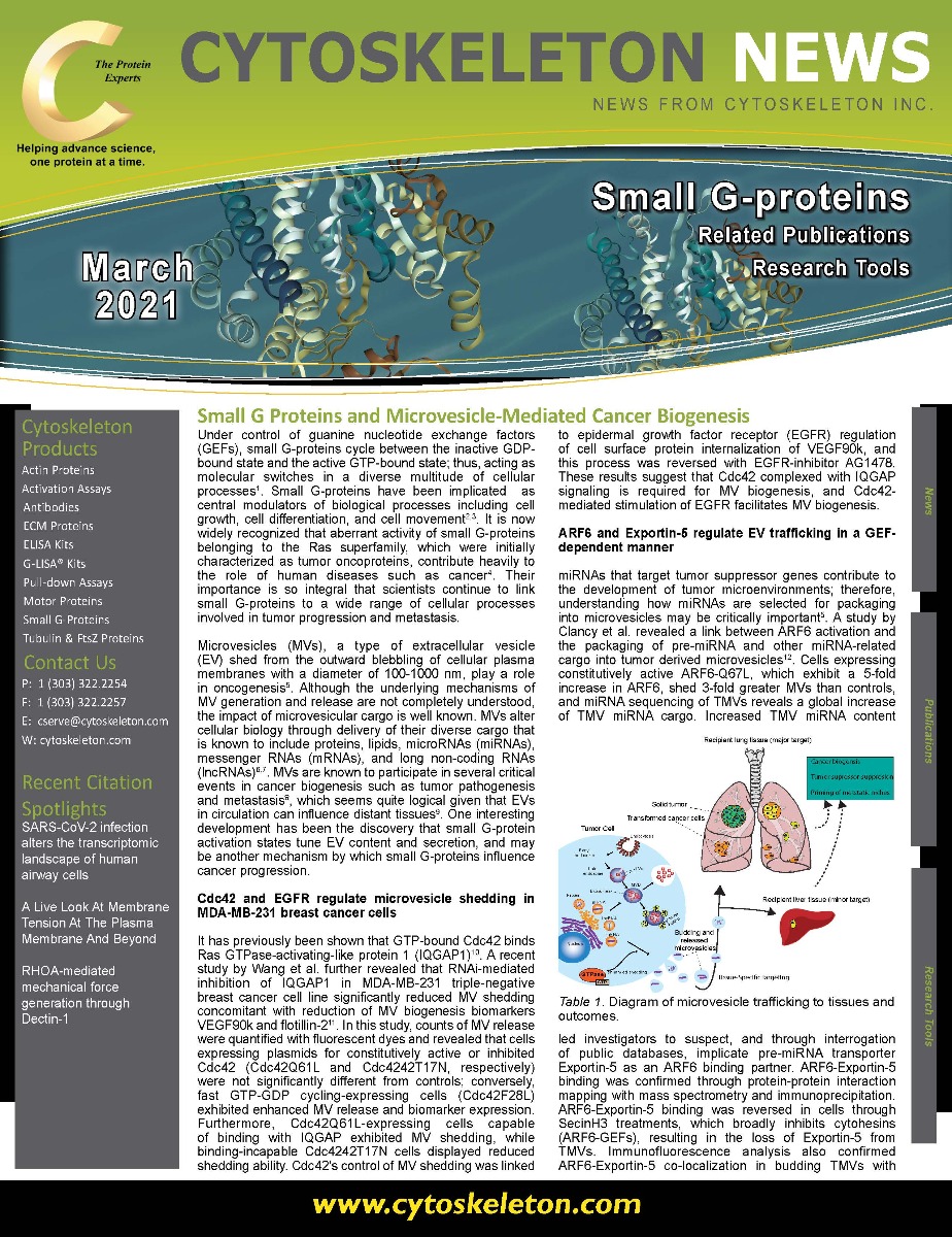

Figure 1. Diagram of microvesicle trafficking to tissues and outcomes.
ARF6 and Exportin-5 regulate EV trafficking in a GEF-dependent manner
miRNAs that target tumor suppressor genes contribute to the development of tumor microenvironments; therefore, understanding how miRNAs are selected for packaging into microvesicles may be critically important5. A study by Clancy et al. revealed a link between ARF6 activation and the packaging of pre-miRNA and other miRNA-related cargo into tumor derived microvesicles12. Cells expressing constitutively active ARF6-Q67L, which exhibit a 5-fold increase in ARF6, shed 3-fold greater MVs than controls, and miRNA sequencing of TMVs reveals a global increase of TMV miRNA cargo. Increased TMV miRNA content led investigators to suspect, and through interrogation of public databases, implicate pre-miRNA transporter Exportin-5 as an ARF6 binding partner. ARF6-Exportin-5 binding was confirmed through protein-protein interaction mapping with mass spectrometry and immunoprecipitation. ARF6-Exportin-5 binding was reversed in cells through SecinH3 treatments, which broadly inhibits cytohesins (ARF6-GEFs), resulting in the loss of Exportin-5 from TMVs. Immunofluorescence analysis also confirmed ARF6-Exportin-5 co-localization in budding TMVs with continued Exportin-5 detection within isolated tumor derived microvesicles. Investigators utilized public databases to deduce cytohesin 3 (GRP1) as the most likely binding partner for the ARF6-GTP-Exportin-5 complex. siRNA-mediated knockdown confirmed that GRP1 acts in concert with ARF6 and Exportin-5 to traffic pre-miRNA cargo into nascent TMVs. This study highlights the importance of the ARF6-Exportin-5 complex as a regulator of miRNA packaging into TMVs.
RalA and RalB control tumor volume and package EVs for tissue-specific metastatic niche formation
Ghoroghi et al. sought to further clarify how RalA and RalB GTPases can tune both the content of TMVs and rate of secretion. Interestingly, it was shown that shRNA-mediated knockdown of RalA or RalB, could significantly reduce the amount of EVs secreted in both human cancer cells lines and the nematode C. elegans; illustrating that EV secretion under control of GTPases is evolutionarily conserved5. Confocal quantification confirmed a concomitant and significant reduction in the number of multivesicular bodies (MVBs), which contain exosomes prior to release; thereby, suggesting that MVBs are under the control of RalA and RalB expression. To determine how GTPases can influence MVB homeostasis investigators focused on phosopholipase Ds (PLDs) which are known to impact cancer progression, exosome secretion, and are targets of RalA and RalB. Suppression of PLD1 through chemical inhibitor CAY10594 in PLD expressing 4T1 cells led to a recapitulation of the suppressed EV secretion and MVB counts seen by RalA/B knockdown. Automated examination of RalA/B protein levels by immunohistochemistry in breast cancer patient-tumors revealed significant overexpression. Xenografting small hairpin RNA (shRNA)-mediated RalA/B-depleted cells into Balb/c mice mammary ducts revealed that while shRalA depletion significantly increased tumor volume, shRalB depletion resulted in tumors with smaller volume than shControl. Intriguingly, although RalA/B depletion increased tumor volume it appeared to reduce metastasis, with shControl-grafted mice exhibiting the highest metastatic foci in serially-sectioned lung tissue.
Functional assays determined that EV content had vascular permeability-enhancing properties that disrupted endothelial cell adherent and tight junctions that could be reversed by small hairpin depletion. EVs isolated from control 4T1 cells were serially injected intra-orbitally into mice to prime metastatic niches and resulted in predominant localization in the lungs that was not observed when EVs were isolated from shRalA/B cells indicating that RalA/B prime EVs to target lung tissues . Interrogation of the EV content with mass spectroscopy revealed differentially expressed proteins found in RalA/B EVs but not shRalA/B EVs, including CD146 and MCAM. Treatment of RalA/B isolated EVs with anti-CD146 and anti-MCAM antibodies abolished lung targeting and pre-metastatic niche formation suggesting that Ral/B-mediating tuning predisposes EVs to enhance metastatic niche formation specifically in lung tissues.
Summary
Altogether these data present an exciting new perspective for small G-protein research. Perturbation of GTPases belonging to the RAS superfamily can modulate the content and quantity of cancer-propagating EVs. Importantly, these works also identified small G-protein accomplices, implicating GEFs and scaffolding proteins, required for microvesicle packaging complexes or downstream effectors which may be important for the development of future druggable targets and improving our understanding of cancer biogenesis. Cytoskeleton has an array of small G-protein tools and membrane probes to aide investigators as they work to define the critical role that small GTPases play in regulating TMVs.
References
- Nestler, E. J. & Duman, R. S. Small G Proteins. (1999).
- Bos, J. L., Rehmann, H. & Wittinghofer, A. GEFs and GAPs: Critical Elements in the Control of Small G Proteins. Cell vol. 129 865–877 (2007).
- Csépányi-Kömi, R., Lévay, M. & Ligeti, E. Small G proteins and their regulators in cellular signalling. Molecular and Cellular Endocrinology vol. 353 10–20 (2012).
- Colicelli, J. Human RAS superfamily proteins and related GTPases. Science’s STKE : signal transduction knowledge environment vol. 2004 RE13 (2004).
- Ghoroghi, S. et al. Ral GTPases promote breast cancer metastasis by controlling biogenesis and organ targeting of exosomes. Elife 10, 1–29 (2021).
- Kelemen, E., Danis, J., Göblös, A., Bata-Csörgő, Z. & Széll, M. Exosomal long non-coding RNAs as biomarkers in human diseases. Electron. J. Int. Fed. Clin. Chem. Lab. Med. 30, 224–236 (2019).
- D’Asti, E. et al. Oncogenic extracellular vesicles in brain tumor progression. Front. Physiol. 3 JUL, 1–15 (2012).
- Wang, T. et al. Hypoxia-inducible factors and RAB22A mediate formation of microvesicles that stimulate breast cancer invasion and metastasis. Proc. Natl. Acad. Sci. U. S. A. 111, (2014).
- D’Souza-Schorey Crislyn, C. & Clancy, J. W. Tumor-derived microvesicles: Shedding light on novel microenvironment modulators and prospective cancer biomarkers. Genes Dev. 26, 1287–1299 (2012).
- Ratajczak, M. Z. & Ratajczak, J. Extracellular microvesicles/exosomes: discovery, disbelief, acceptance, and the future? Leukemia vol. 34 3126–3135 (2020).
- Wang, J. et al. Cdc42 functions as a regulatory node for tumour-derived microvesicle biogenesis. J. Extracell. Vesicles 10, (2021).
- Clancy, J. W., Zhang, Y., Sheehan, C. & D’Souza-Schorey, C. An ARF6–Exportin-5 axis delivers pre-miRNA cargo to tumour microvesicles. Nat. Cell Biol. 21, 856–866 (2019).
Related Products
Bead pull-down Activation Assays
Arf1 Activation Assay Biochem Kit (Cat. # BK032-S)
Arf6 Activation Assay Biochem Kit (Cat. # BK033-S)
GGA3-PBD Beads (Arf1 + Arf6) (Cat. # GGA07)
Cdc42 Activation Assay Biochem Kit (Cat. # BK034)
Rac + Cdc42 Activation Assay Biochem Kit (Cat. # PAK02)
Rac1 Activation Assay Biochem Kit (Cat. # BK035)
Ras Activation Assay Biochem Kit (Cat. # BK008)
Rhotekin RBD beads (RhoA/B/C) (Cat. # RT02)
RhoA Activation Assay Biochem Kit (Cat. # BK036)
RhoA/Rac1/Cdc42 Activation Assay Combo Biochem Kit (Cat. # BK030)
Actin Biochem Kits
Arf1 G-LISA™ Activation Assay Kit (Colorimetric format) (Cat. # BK132)
Arf6 G-LISA™ Activation Assay Kit (Colorimetric format) (Cat. # BK133)
Cdc42 G-LISA™ Activation Assay Kit (Colorimetric format) (Cat. # BK127)
Rac1 G-LISA™ Activation Assay Kit (Luminescence format) (Cat. # BK126)
Rac1 G-LISA™ Activation Assay Kit (Colorimetric format) (Cat. # BK128)
RalA G-LISA™ Activation Assay Kit (Colorimetric format) (Cat. # BK129)
RhoA G-LISA™ Activation Assay (Luminescence format) (Cat. # BK121)
RhoA G-LISA™ Activation Assay Kit (Colorimetric format) (Cat. # BK124)
RhoA / Rac1/ Cdc42 G-LISA™ Activation Assay Bundle 3 kits (24 assays per kit) (Cat. # BK135)
Rac1,2,3 G-LISA™ Activation Assay (Colorimetric format) (Cat. # BK125)
Rac1 G-LISA™ Activation Assay (Luminescence format) (Cat. # BK126)
Rac1 G-LISA™ Activation Assay Kit (Colorimetric Based) (Cat. # BK128)
Ras G-LISA™ Activation Assay Kit (Colorimetric Based) (Cat. # BK131)