October 2020 Newsletter: Mitotic Spindle - New Tools for Visualization
Mitotic Spindle - New Tools for Visualization
Mitotic Spindle Background
Cell division is a requisite task for all living things. A critical part of this process is the formation of the mitotic spindle which is the machinery that actively and effectively divides chromosomes into their respective daughter cells during mitosis. The overall morphology of the mitotic spindle is well-defined with kinetochore microtubule bundles ( aka: kinectochore fibers) extending from the poles of centrosomes and attaching to specialized attachment sites on sister chromatids called kinetochores to achieve the processes that occur in metaphase and anaphase. Conversely, the molecular mechanisms regulating mitotic spindle function have been much more challenging to decipher for several reasons: 1. it is extremely large and complex, often comprised of hundreds to thousands of microtubules, 2. it is one of the most dynamic cytoskeletal structures, and 3. there are a multitude of microtubule associated proteins (MAPs) that participate in the formation and function of this structure(reviewed in (1, 2)). Improved microtubule visualization tools will allow investigators to better understand the mechanisms that regulate mitotic spindle spatiotemporal organization, unique spindle forces, and spindle interaction with kinectochore complexes.
Microtubules (MTs) are formed by the addition of ab-heterodimers of tubulin. A striking property of MTs are their dynamic instability which is critical for its many functions in the cell including its role in mitotic spindle formation and mitosis. Studying MT function is challenging due to its dynamic properties; however, in vitro approaches such as turbidity measurements, DIC microscopy, and fluorescently labeled tubulins in combination with TIRF microscopy have allowed scientists to better understand MT growth speed, shrinkage speed, catastrophe rate, and rescue rate(reviewed in (3)). Investigating MTs in cells presents its own challenges, because labeling tubulin in cells can affect binding properties, associated protein interaction, and dynamic regulation. For example, in cells standard overexpression of fluorescent proteins fused to tubulin produces steric hinderance and often results in high overall background signals. To circumvent this issue and aide in dynamic investigation, scientists use a “speckling” approach where only some of the tubulin is fluorescently labeled either by microinjecting a fixed amount of fluorescently labeled tubulin into the cell, or expressing GFP on a specific isoform of tubulin. Recently, a new fluorescently labeled tubulin specific probe, called SiR tubulin, has gained significant interest due to its specificity, cell-permeability, and fluorogenic properties(4). Following are three examples of how SiR tubulin technology has enhanced investigation of the mitotic spindle.
Specific forces are required in mitosis for the congression of chromosomes to the metaphase plate and their separation in anaphase. Several studies have determined that forces generated in the mitotic spindle are generated by motor proteins. Interestingly, Novak M et al. recently discovered that the motor protein kinesin-5, in addition to generating force, also creates torque in the mitotic spindle(5). Stimulated emission depletion (STED) super-resolution microscopy was used in combination with SiR tubulin to determine the shapes of MT bundles in metaphase spindles, which allowed them to conclude that MTs are arranged in bundles exhibiting a variety of shapes including curved and S shapes. They then utilized PRC1-GFP fusion protein to label the kinectochore fibers in fixed cells and performed confocal microscopy, which allowed them to re-create the z-stacked kinectochore fibers and determine that the mitotic spindle is a chiral object with left-handed helicity of the MT bundles. Interestingly when kinesin-5 was inactivated with S-trityl-L-cysteine the bundles had reduced left-handed helicity. It will be interesting to determine the role that torque forces play in mitotic spindle formation and function.
Spatiotemporal Organization of the Mitotic Spindle
The formation of the mitotic spindle is coordinated by cytoskeletal networks, molecular motors, and the nucleus. Recently, Nunes et al. investigated this processes in a spatiotemporal fashion and identified several key mechanisms that ensure efficient spindle assembly(6). Micropatterning with high-resolution imaging and 3D cellular reconstruction was used to investigate this complex process, and they determined that centrosomes are reoriented during prophase so that it is positioned on the shortest nuclear axis when the nuclear envelope is broken down, and understanding this positioning is critical because it can affect mitotic fidelity. Furthermore, they found that Arp 2/3 and Dyenin are key regulators of this centrosome movement. SiR tubulin was essential in their studies to determine if spindle assembly checkpoint was impacted by centrosome positioning, and they found that centrosomes positioned at the shortest nuclear axis removed Mad2 faster and produced timely progression through mitosis.
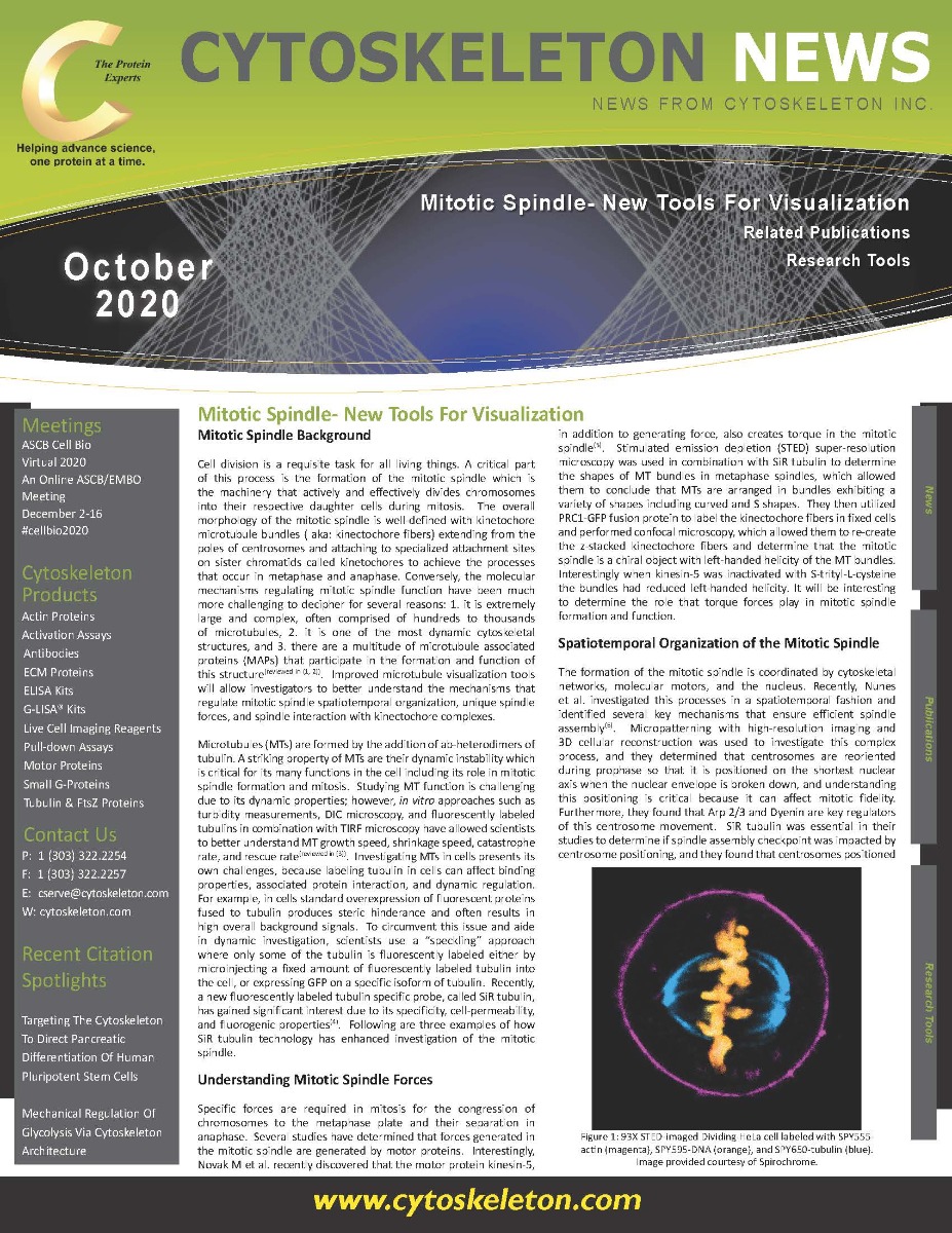
Figure 1: 93X STED-imaged Dividing HeLa cell labeled with SPY555-actin (magenta), SPY595-DNA (orange), and SPY650-tubulin (blue). Image provided courtesy of Spirochrome.
Phosphorylation of Kinetochore Proteins to Regulate Kinetochore Coupling
Kinetochores are key structures in mitosis that actively bind kinetochore fibers during mitosis. Long et al. sought to decipher mechanisms that regulate kinectochore interaction with polymerizing and depolymerizing MT bundles during metaphase(7). The Hec1 protein, a key component of the kinectochore-MT attachment interface, has tails that are phosphorylated by aurora B. Interestingly, the investigators found that the Hec1 tail phosphorylation state alters its ability to interact with polymerizing microtubule bundles by tuning friction along polymerizing microtubules while not affecting Hec1’s grip on depolymerizing microtubules. SiR tubulin was used in primary studies to define if Hec1 phospho-mimetics altered its ability to interact with MTs.
Summary and Future Investigation
These studies highlight how SiR tubulin probes have played a critical role, in combination with established tools, to better define mitotic spindle formation and function. Just as importantly, these studies highlight microtubule associated proteins (MAPs) that control critical mechanisms in the mitotic spindle. MAPs were first identified by Greengard and colleagues in 1975(8) and since then several proteomic and functional studies have identified more than 200 of these proteins involved in mitotic spindle formation and function(reviewed in (1)). As scientists continue to dive deeper into understanding the role of MAPs in the dynamic regulation of the mitotic spindle using super-resolution microscopy, better live cell imaging tools for MAPs are needed. GFP fusion proteins are an established approach; however, phototoxicity and signal-to-noise is an issue with GFP and can be a limiting factor especially for single molecule and super-resolution microscopy where fluorescent probes are preferred. An alternative fusion-protein system is the SNAP tag labeling system, which allow investigators to specifically label their proteins with an array of benzylguanine (BG) conjugated fluorophores(9). This allows for the potential labeling of robust fluorescent dyes; nevertheless, not all BG-probes are created equally and one recent study showed that many of these probes result in rapid photobleaching and nonspecific staining(10). Cytoskeleton is proud to introduce the BG-SiR and BG-SPY dyes which are composed of BG conjugated to the cell permeable, fluorogenic dyes used to make the highly popular SiR and SPY tubulin probes. Importantly, these probes can be used in combination for super-resolution microscopy (Figure 1). This new toolset provides a great mechanism to label both tubulin and MAPs with well-established cell-permeable and fluorogenic probes for mitotic spindle investigation.
References
Petry S. Mechanisms of Mitotic Spindle Assembly. Annu Rev Biochem. 2016;85:659-83.
McIntosh JR. Mitosis. Cold Spring Harb Perspect Biol. 2016;8(9).
Lukinavicius G, Reymond L, D'Este E, Masharina A, Gottfert F, Ta H, et al. Fluorogenic probes for live-cell imaging of the cytoskeleton. Nat Methods. 2014;11(7):731-3.
Novak M, Polak B, Simunic J, Boban Z, Kuzmic B, Thomae AW, et al. The mitotic spindle is chiral due to torques within microtubule bundles. Nat Commun. 2018;9(1):3571.
Nunes V, Dantas M, Castro D, Vitiello E, Wang I, Carpi N, et al. Centrosome-nuclear axis repositioning drives the assembly of a bipolar spindle scaffold to ensure mitotic fidelity. Mol Biol Cell. 2020;31(16):1675-90.
Long AF, Udy DB, Dumont S. Hec1 Tail Phosphorylation Differentially Regulates Mammalian Kinetochore Coupling to Polymerizing and Depolymerizing Microtubules. Curr Biol. 2017;27(11):1692-9 e3.
Sloboda RD, Rudolph SA, Rosenbaum JL, Greengard P. Cyclic AMP-dependent endogenous phosphorylation of a microtubule-associated protein. Proc Natl Acad Sci U S A. 1975;72(1):177-81.
Keppler A, Gendreizig S, Gronemeyer T, Pick H, Vogel H, Johnsson K. A general method for the covalent labeling of fusion proteins with small molecules in vivo. Nat Biotechnol. 2003;21(1):86-9.
Bosch PJ, Correa IR, Jr., Sonntag MH, Ibach J, Brunsveld L, Kanger JS, et al. Evaluation of fluorophores to label SNAP-tag fused proteins for multicolor single-molecule tracking microscopy in live cells. Biophys J. 2014;107(4):803-14.
, SPY595-DNA (orange), and SPY650-tubulin (blue). Image provided courtesy of Spirochrome.")