Inflammatory Bowel Disease
Inflammatory bowel disease (IBD) is a group of gastrointestinal disorders characterized by severe inflammation and includes ulcerative colitis (UC) and Crohn's disease. IBD is thought to result from a combination of several factors including an inflammatory response to commensal microbes, dysregulation of the mucosal immune response, and a loss of epithelial barrier integrity (reviewed in [1, 2]). The intestinal epithelial barrier is a monolayer of epithelial cells that form a mechanical link to one another via adhesive structures such as tight junctions and adherens junctions. These adhesive, apical structures are comprised of adhesive and scaffolding proteins that are bound to cytoskeletal structures like actin filaments[2].
The integrity of the epithelial barrier is critical for the proper absorption of key nutrients and protection from external pathogens in the gastrointestinal tract; conversely, disruption of the intestinal epithelial layer results in a leaky barrier that can manifest into IBD. Researchers have sought to understand the factors that contribute to IBD such as how bacterial factors affect apical junctional complexes, immune cell activation, and cytoskeletal structures. Below, we look at recently identified mechanisms that control the actin cytoskeleton in IBD.
Actin Cytoskeleton Controls Epithelial Barrier Integrity
Critical proteins within the apical junction complexes (AJC) are deregulated in IBD pathogenesis (reviewed in [1]); additionally, recent mass spectrometry proteomic studies of IBD from human samples as well as animal models identified measurable changes in actin expressed in the inflamed gut [3, 4]. These studies align with the overarching hypothesis that actin plays a role in the AJC and overall barrier function; furthermore, it supports earlier findings that disruption of actin with cytoskeleton-disrupting toxins, inflammatory signaling molecules, or bacterial pathogens leads to loss of apical junctions that coincides with actin filament breakdown (reviewed in [2]). A recent study by the Ivanov group sought to define the role of b-actin in IBD in an in vivo model and therefore utilized a mouse model where b-actin was specifically knocked out from intestinal epithelial cells to further define actin’s role in the epithelial barrier [5]. They found that these mice did not have overt gastrointestinal abnormalities, which they ascribed to a potential compensation by g-actin. However, they did note that these mice showed increased intestinal permeability and a more pronounced effect to dextran sodium sulfate, a chemical inducer of colitis. These data provide in vivo evidence that b-actin is important for proper epithelial barrier function.
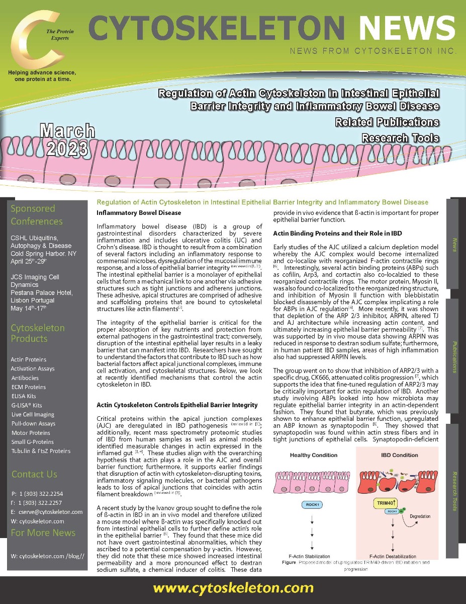

Figure. Proposed model of upregulated TRIM40-driven IBD initiation and
progression
Actin Binding Proteins and their Role in IBD
Early studies of the AJC utilized a calcium depletion model whereby the AJC complex would become internalized and co-localize with reorganized F-actin contractile rings [6]. Interestingly, several actin binding proteins (ABPs) such as cofilin, Arp3, and cortactin also co-localzied to these reorganized contractile rings. The motor protein, Myosin II, was also found co-localized to the reorganized ring structure, and inhibition of Myosin II function with blebbistatin blocked disassembly of the AJC complex implicating a role for ABPs in AJC regulation [6]. More recently, it was shown that depletion of the ARP 2/3 inhibitor, ARPIN, altered TJ and AJ architecture while increasing actin content, and ultimately increasing epithelial barrier permeability [7]. This was supported by in vivo mouse data showing ARPIN was reduced in response to dextran sodium sulfate; furthermore, in human patient IBD samples, areas of high inflammation also had suppressed ARPIN levels. The group went on to show that inhibition of ARP2/3 with a specific drug, CK666, attenuated colitis progression [7], which supports the idea that fine-tuned regulation of ARP2/3 may be critically important for actin regulation of IBD. Another study involving ABPs looked into how microbiota may regulate epithelial barrier integrity in an actin-dependent fashion. They found that butyrate,a product of specific bacteria in the microbiome, which was previously shown to enhance epithelial barrier function, upregulated an ABP known as synaptopodin [8]. They showed that synaptopodin was found within actin stress fibers and in tight junctions of epithelial cells. Synaptopodin-deficient mice had exacerbated colitis phenotype in response to dextran sodium sulfate sodium[8]. This study identified a critical mechanism by which microbiota regulates intestinal barrier function through the regulation of actin via ABPs.
Other Mechanisms that Regulate Actin in Epithelial Barrier Regulation
It was recently shown that a subset of patients with IBD had increased levels of tripartite motif-containing protein 40 (TRIM40), which is an ubiquitin E3 ligase that is known to target ROCK1 [9]. Depletion of ROCK1 by TRIM40 suppresses key signaling mechanisms that normally promote actin formation and ROCK1/Rho signaling[9]; a key signaling pathway that has been implicated in AJC disassembly and reduced epithelial barrier function (reviewed in [2]). In the study, TRIM40 deficient mice were highly resistant to chemical-induced colitis[9] further supporting the hypothesis that TRIM40 is a critical regulatory mechanism in IBD progression. Tsukita et al. investigated the role of Liquid-liquid phase separation (LLPS) is a mechanism that enables for the rapid enhancement in concentration of a given protein and has been shown to be important for dynamic cellular mechanisms. A recent study identified zonula occludens-1, a protein within the AJC, concentration is modified by LLPS and is important for epithelial barrier formation [10]. In another study, Tsukita et al. found that microtubules facilitate LLPS of a novel microtubule-binding protein cordon bleu [11]. Once in the LLPS at the apical junction complex, cordon bleu, which is a novel actin nucleator, promoted actin assembly to regulate the epithelial barrier. This study identified a novel crosstalk mechanism between microtubules and actin that was important for fine-tuning epithelial barrier function.
Summary and Future Perspectives
Studies show that approximately 6.8 million people worldwide suffer from IBD, and it’s now understood that IBD is a major risk factor leading to the development of colorectal cancer (CRC) [12]. In a study performed by Kanaan et al. (2010), normal, dysplastic, and cancerous tissue was obtained from three patients with IBD-associated CRC. After mRNA extraction, oligonucleotide arrays were performed to identify genes that were differentially expressed between the three tissues. They observed that genes involved in the actin cytoskeleton were significantly dysregulated between the three tissues, therefore linking the actin-cytoskeleton pathway to the progression of normal colonic mucosa, via dysplasia, to IBD-associated CRC [13]. Understanding the mechanisms by which epithelial barrier function is dysregulated including through regulation of AJC and cytoskeletal actin structures may allow for the prevention of IBD.
References
- Chelakkot, C., J. Ghim, and S.H. Ryu, Mechanisms regulating intestinal barrier integrity and its pathological implications. Exp Mol Med, 2018. 50(8): p. 1-9.
- Ivanov, A.I., C.A. Parkos, and A. Nusrat, Cytoskeletal regulation of epithelial barrier function during inflammation. Am J Pathol, 2010. 177(2): p. 512-24.
- Moriggi, M., et al., Contribution of Extracellular Matrix and Signal Mechanotransduction to Epithelial Cell Damage in Inflammatory Bowel Disease Patients: A Proteomic Study. Proteomics, 2017. 17(23-24).
- Cooney, J.M., et al., A combined omics approach to evaluate the effects of dietary curcumin on colon inflammation in the Mdr1a(-/-) mouse model of inflammatory bowel disease. J Nutr Biochem, 2016. 27: p. 181-92.
- Lechuga, S., et al., Loss of beta-Cytoplasmic Actin in the Intestinal Epithelium Increases Gut Barrier Permeability in vivo and Exaggerates the Severity of Experimental Colitis. Front Cell Dev Biol, 2020. 8: p. 588836.
- Ivanov, A.I., et al., Role for actin filament turnover and a myosin II motor in cytoskeleton-driven disassembly of the epithelial apical junctional complex. Mol Biol Cell, 2004. 15(6): p. 2639-51.
- Chanez-Paredes, S., et al., The Arp2/3 Inhibitory Protein Arpin Is Required for Intestinal Epithelial Barrier Integrity. Front Cell Dev Biol, 2021. 9: p. 625719.
- Wang, R.X., et al., Microbiota-derived butyrate dynamically regulates intestinal homeostasis through regulation of actin-associated protein synaptopodin. Proc Natl Acad Sci U S A, 2020. 117(21): p. 11648-11657.
- Kang, S., et al., TRIM40 is a pathogenic driver of inflammatory bowel disease subverting intestinal barrier integrity. Nat Commun, 2023. 14(1): p. 700.
- Beutel, O., et al., Phase Separation of Zonula Occludens Proteins Drives Formation of Tight Junctions. Cell, 2019. 179(4): p. 923-936 e11.
- Tsukita, K., et al., Phase separation of an actin nucleator by junctional microtubules regulates epithelial function. Sci Adv, 2023. 9(7): p. eadf6358.
- Stidham, R.W. and P.D.R. Higgins, Colorectal Cancer in Inflammatory Bowel Disease. Clin Colon Rectal Surg, 2018. 31(3): p. 168-178.
- Kanaan, Z., et al., The actin-cytoskeleton pathway and its potential role in inflammatory bowel disease-associated human colorectal cancer. Genet Test Mol Biomarkers, 2010. 14(3): p. 347-53.