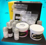
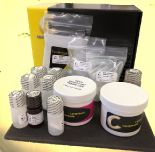
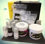
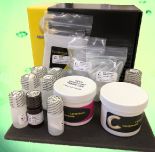
Signal-Seeker™ Phosphotyrosine Detection Kit (10 assay)
The Signal-Seeker™ line of produts have been developed to allow simple analysis of key regulatory protein modifications by specialists and non-specialists alike. The comprehensive Signal-Seeker™ kits provide an affinity bead system to isolate and enrich modified proteins from any given cell or tissue lysate. The enriched protein population is then analyzed by standard western blot procedures using a primary antibody to the target protein.
Product Uses Include
- Investigate transient regulatory mechanisms
- Measure signalling events of multiple pathway member proteins
- Discover new modifications of your protein of interest
- Gain insight into regulatory mechanisms
- Measure endogenous or transiently expressed protein signalling events
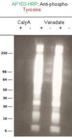
Validation Data: Phosphotyrosine Detection Kit White Paper
Kit contents
The Phosphotyrosine kit contains the following components:
| Lysis and protein quantitation step | IP and pre-clear step | Wash step | Elution step | Western step |
BlastR™ Lysis Buffer BlastR™ Dilution Buffer BlastR™ Filters Protease Inhibitor Cocktail Tyrosine Phosphatase inhibitor Cocktail Precision Red Protein Assay Reagent | Phosphotyrosine Affinity Beads IgG Control Beads
| BlastR™ Wash Buffer
| Spin Columns Bead Elution Buffer
| Chemiluminescent Reagent A Chemiluminescent Reagent B Anti-phosphotyrosine-HRP antibody
|
Example results
There are many applications for these kits (see next section below), here we describe an interesting example:
Application 1: Investigate highly transient regulation events
Temporal changes in Rac1 tyrosyl phosphorylation were examined relative to Rac1 activation in response to EGF treatment. HeLa cells were grown to 50% confluency and treated for varying amounts of time (0.5 min-30 min) with 50 ng/ml EGF. The Rac1 activation profile (performed using kit BK035) showed a classical activation pattern in which Rac1 activation peaked rapidly after EGF treatment (0.5 min), thereafter falling to basal levels. The phosphotyrosine Rac1 profile (performed using kit BK160) revealed a modification pattern in which Rac1 tyrosyl phosphorylation was rapidly reduced from basal levels after EGF treatment (0.5-1 min) thereafter returning to basal levels. Each lane represents immunoprecipitated phosphotyrosine proteins from 1000 μg of cell lysate. The figures shown to he right are representative of at least 5 repeats. Lane IN represents 10 μg of input lysate.
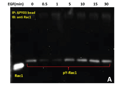
Figure 1: Serum starved HeLa cells were either left untreated or treated with EGF (50ng/ml) for specific times as indicated. Cells were lysed in RIPA buffer.APY03 beads were used to immunoprecipitate pY proteins from 1 mg of total cell lysate from each condition. Samples were resolved by SDS-PAGE and immunoblotted with anti-Rac1 antibody to detect tyrosyl phosphorylated-Rac1 from the immunoprecipitated pY protein population
Other experiments that could be attempted in this area of research include:
• Pharmacological investigation of kinases / phosphatases involved in Rac1 tyrosyl phosphorylation.
• Investigation of the relationship between Rac1 activation and tyrosyl phosphorylation under a variety of different growth factors or drug treatments.
• Examine the interaction of phosphotyrosine Rac1 with effectors.
• Examine the potential crosstalk between phosphotyrosine Rac1 and other PTMs that modify this protein.
For more information contact: signalseeker@cytoskeleton.com
Associated Products:
Signal-Seeker™ Ubiquitination Detection Kit (Cat. # BK161)
Signal-Seeker™ SUMOylation 2/3 Detection Kit (Cat. # BK162)
Signal-Seeker™: BlastR™ Rapid Lysate Prep Kit (Cat. # BLR01)
Signal-Seeker™ Phosphotyrosine Affinity Beads (Cat.# APY03-beads)
Signal-Seeker™: PTMtrue™ Phosphotyrosine Antibody (Cat.# APY03)
Visit our Signal-Seeker™ Tech Tips and FAQs page for technical tips and frequently asked questions regarding this and other Signal-Seeker™ products click here
If you have any questions concerning this product, please contact our Technical Service department at tservice@cytoskeleton.com


