SUMO-2/3 Antibody Mouse Monoclonal (Clone 12F3)

Host/Isotype
Mouse / IgG2a-kappa
Clone
12F3
Species Reactivity
Broad Reactivity
Validation Data
White Paper
Antibodypedia
RRID
AB_2884967
Anti-SUMO2/3 antibody is a SUMO-2/3 mouse monoclonal antibody that is part of the Signal-Seeker™ product line. Anti-SUMO-2/3 mouse monoclonal antibody was raised against full-length recombinant SUMO-2 protein (Uniprot: P61956) combined with a proprietary mix of peptides that include CQIRFRFDGQPINE. The antibody has been shown to recognize a wide range of SUMO-2/3-targeted proteins in HeLa cell lysate (Fig. 1B) and to detect sub-nanogram amounts of recombinant SUMO-2 (Fig. 1A). Epitope mapping has identified that the antibody recognizes a sequence/structure within the peptide CQIRFRFDGQPINE. The peptide sequence is conserved in mammals, birds, and amphibians, giving the antibody broad species reactivity. ASM23 is purified by Protein G affinity chromatography and is supplied as a lyophilized white powder.
Each Lot of antibody is quality controlled to provide a high batch to batch consistency. The Lot specific µg per tube can be found in the Lot specific COA documents.

Validated Applications
Figure 1: Western Blot using SUMO-2/3 Antibody
12F3 was used for immunoblotting (1:500 dilution) following the recommended Western blot protocol (see below). Figure 1A: Titration of recombinant SUMO-2 (40-0.6 ng) and SUMO-1 (800 ng). SUMO-2 was detected down to 0.6 ng while SUMO-1 was not detected at 800 ng. Figure 1B: Induction of SUMOylation by heat shock and reduction of SUMOylation by SUMO-2 shRNA knockdown. Cell lysates were prepared from HeLa cells: Lane 2: Heat Shock treated (43°C for 10min), Lane 3: untreated, Lane 4: shRNA SUMO-2 knock down. 20 µg of HeLa cell lysates were used for each sample. Lane 1: position of molecular weight markers. Figure 1C: Specificity of SUMO-2 knockdown signal. Lane 1: parental HeLa cell lysates, Lane 2: SUMO-2 shRNA control lysates, Lane 3: SUMO-1 shRNA knock-down cell lysates, Lane 4: SUMO-2 shRNA knock-down cell lysates. Arrow head indicates free SUMO-2/3. To see the full Western blot protocol, see the product datasheet.
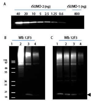
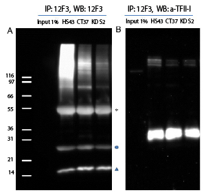
Figure 2: Immunoprecipitation using SUMO-2/3 Antibody
Denatured cell lysates were prepared from HS43, CT37 and KD S212. 1mg of lysate was used for the immunoprecipitation of SUMO-2/3 conjugates. IP experiments were performed by the protocol presented in IP Method. Western blots of immunoprecipitated proteins were developed using 12F3 (A) or anti-TFII-I antibody (B). (A) Star (*) and circle (o) indicate heavy and light chains of antibodies. Unconjugated free SUMO is denoted by triangle. (B) Unconjugated TFII-I is visible near 120 kDa. Multiple bands indicate that TFII-I is SUMOylated by several SUMO-2/3 proteins. TFII-I has previously been reported to be a target for Sumoylation . To see the full Immunoprecipitation protocol, see the product datasheet.
HeLa cells were stained and visualized by confocal fluorescence microscopy as described in the IF method below. The cells were stained against α/β-tubulin (sheep anti-tubulin Ab, Cat# ATN02, green) and SUMO-2/3 (12F3, red). DNA was stained with DAPI. Mitotic cells in metaphase were imaged with a Zeiss LSM 780 confocal microscope (1.4 NA 63X objective). Detection of SUMO 2/3 at chromosomes can be observed during mitosis as has been previously reported10. To see the full Immunofluorescence protocol, see the product datasheet.
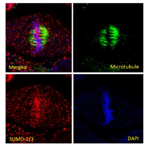
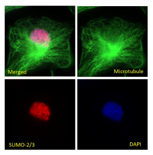
Figure 4: Immunofluorescence of HeLa cells in interphase with SUMO-2/3 Antibody
HeLa cells were stained and visualized by widefield fluorescence microscopy as described in the IF method below. The cells were stained against α/β-tubulin (sheep anti-tubulin Ab, Cat# ATN02, green) and SUMO-2/3 (12F3, red). DNA was stained with DAPI. Cells in interphase were imaged with a Zeiss Axio Observer.Z1 microscope (1.4 NA 63X objective). PML nuclear bodies (nuclear dots) were visible in SUMO-2/3 staining as has been previously reported11. To see the full Immunofluorescence protocol, see the product datasheet.
For more information contact: signalseeker@cytoskeleton.com
Associated Products:
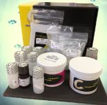
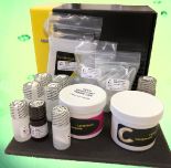
Signal-Seeker™ SUMOylation 2/3 Detection Kit (Cat. # BK162)
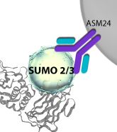
Signal-Seeker™ SUMOylation 2/3 Affinity Beads (Cat.# ASM24-beads)
Signal-Seeker™: BlastR™ Rapid Lysate Prep Kit (Cat. # BLR01)
For product Datasheets and MSDSs please click on the PDF links below.
Sample Size Datasheet (Cat. ASM23-S): ![]()
Certificate of Analysis: Lot 011
Visit our Signal-Seeker™ Tech Tips and FAQs page for technical tips and frequently asked questions regarding this and other Signal-Seeker™ products click here
If you have any questions concerning this product, please contact our Technical Service department at tservice@cytoskeleton.com