Why Does K-Ras Display Oncogenic Specificity?
Ras GTPases regulate cell proliferation pathways, making them important molecules in oncogenesis and cancer cell migration and invasion1-4. The four isoforms of Ras, H-Ras, N-Ras, K-Ras4A, and K-Ras4B (due to alternative splicing), were identified over 30 years ago for their oncogenic activation in human tumors1,2. Activating Ras mutations are single amino acid substitutions (e.g., G12C, G12V, G12D) and have been identified in approximately 30% of all human cancers5-7.
The same signaling pathways activate all Ras isoforms via guanine exchange factor (GEF)-mediated exchange of GDP for GTP, followed by binding to the same effector proteins. However, Ras oncogenic isoforms are differentially expressed aacross different cancers with oncogenic specificity significantly favoring K-Ras2,8-11. Indeed, K-Ras is the most common mutated Ras isoform (86% of all Ras mutations) and is correlated with over 21% of human cancers5. In particular, K-Ras is the predominant or exclusive Ras gene mutated in three of the top four cancers with the highest mortality rates in the US: lung, colon, and pancreatic cancers5. In most instances, the K-Ras4B is the primary isoform mutated in K-Ras-associated cancers8. This newsletter discusses potential explanations for the biological basis of K-Ras’s oncogenic specificity.
Membrane Subdomain Localization of Ras
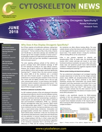
For GEF-mediated activation of Ras and subsequent interaction with downstream effectors, Ras first must be trafficked, inserted, and anchored to different subdomains on the inner surface of the plasma membrane (termed nanoclustering). Localization is isoform-specific and determined by each isoform’s particular lipid post-translational modifications (PTMs; e.g., palmitoyl, farnesyl, geranylgeranyl) within the C-terminus region known as the hypervariable region (HVR)2,6,10-12 (Fig. 1). Palmitoylation favors membrane insertion into lipid microdomains such as lipid rafts or liquid-ordered phase membranes. Farnesylation and geranylgeranylation primarily favor membrane insertion into liquid-ordered phase membranes. H-Ras has one farnesyl and two palmitoyls lipid attachments, while N-Ras and K-Ras4A have only one palmitoyl. In contrast, K-Ras4B has only one farnesyl and a charged polybasic HVR (Fig. 1). K-Ras’s unique lipid PTM profile favors acid membranes that are disordered in opposition to the membrane composition that H- and N-Ras favor2,6,10-12. Differences in PTMs and the resulting conformations within the membrane can affect effector binding affinity. The same catalytic domain surface that interacts with the effector protein can also engage with the plasma membrane12. Even in the GTP-bound state, if the catalytic domain is facing the membrane, effector binding affinity can be compromised13.

Figure 1. Primary structure of Ras isoforms. Adapted from ref. # 2.
K-Ras is also uniquely regulated by ubiquitin and phosphorylation PTMs. At steady state, the deubiquitinating enzyme USP17 inhibits wild-type and mutant H- and N-Ras functional membrane localization, but spares K-Ras, both under steady state and epidermal growth factor (EGF)-stimulated conditions14. Phosphorylation also negatively regulates K-Ras4B membrane binding and clustering2,15-17.
Effector Protein Binding and Downstream Pathways
The two predominant physiological and oncological signaling cascades downstream of active Ras are the mitogen-activated protein kinase (MAPK) and phosphoinositide-3-kinase (PI3K) pathways12. Active Ras isoforms anchored to the plasma membrane promote Raf-1 dimerization and activate MAPK. Activated K- and H-Ras nanoclusters recruit Raf-1, but it is only retained within the K-Ras nanoclusters, making K-Ras a more potent activator of Raf-1 than H-Ras18,19. With respect to PI3K, K-Ras is a weaker activator than the other isoforms18, but has the potential to activate PI3K in the absence of receptor tyrosine kinase (RTK) stimulation12,20,21.
K-Ras4B is also specifically required for PDGF-mediated activation of Akt and subsequent enhanced cell migration22. This pathway also requires calmodulin (CaM) and CaM’s role in growth factor-mediated activation of Akt requires K-Ras4B, likely involving the PDGF-stimulated increase in K-Ras/CaM-Ca2+ complex formation22.
Distinct Binding Partners
Researchers have posited that at least one unique K-Ras binding partner exists that mediates its unique oncogenic specificity. Two candidates are the CaM-Ca2+ complex and PDEδ12,20,21. CaM-Ca2+ binds only the K-Ras4B isoform with the HVR farnesyl moiety, docking in a pocket on CaM in a Ca2+-dependent manner20-24. This binding removes K-Ras4B from the membrane24,25. The CaM-Ca2+/K-Ras4B complex also offers a potential pathway by which oncogenic K-Ras4B could activate cell proliferation pathways. Under physiological conditions, Ras-mediated PI3K signaling is only fully engaged in the presence of RTK stimulation. However, CaM-Ca2+ can bind and activate PI3K26, and because only K-Ras4B is capable of binding to CaM-Ca2+, researchers suggest that a K-Ras4B/CaM-Ca2+/PI3K complex can form, which allows the CaM-Ca2+ complex to substitute for normal RTK-mediated activation of the Ras-PI3K signaling cascade12,20,21. In this scenario, oncogenic K-Ras4B mutants could activate cell proliferation pathways in the absence of physiological RTK stimulation12,20,21. In addition, binding of K-Ras to CaM-Ca2+ suppresses non-canonical Wnt/Ca2+ signaling, resulting in facilitation of K-Ras-mediated tumorigenesis27.
Another unique K-Ras4B binding partner is PDEδ, which is responsible for trafficking of K-Ras4B to the membrane, while H-, N-, and K-Ras4A rely upon vesicles12,24,28,29. Thus, availability of PDEδ can regulate K-Ras trafficking and subsequent membrane localization and function.
Conclusions
The question persists – why is there oncogenic bias toward K-Ras? Besides the differences in conformation, localization, and binding partners discussed above, K-Ras might contribute a unique developmental role as suggested by K-Ras4B knock-out animal studies2,30,31. In any case, the clinical oncology data indicate the importance of targeting Ras proteins, and specifically K-Ras4B, to treat the largest percentage of Ras-driven cancers. To better understand the role of all Ras isoforms in physiological and oncological processes, Cytoskeleton offers a variety of Ras-centric proteins and assay kits.
Related Products & Resources
H-Ras protein: His tagged: human wild type (Cat. # RS01-A)
N-Ras Protein, human rec., wild type (Cat. # CS-RS02)
K-Ras4B Protein, human rec., wild-type (Cat. # CS-RS03)
K-Ras4B Protein, human rec., G12V mutant (Cat. # CS-RS04)
SOS1 Ras GEF Domain Protein, GEF for H-, K- or N-Ras (Cat. # CS-GE02)
RasGRF1 GEF Protein (Cdc25 Exchange Domain, aa1038-1270, MBP tag) (Cat. # CS-GE03)
Ras Pull-down Activation Assay Biochem Kit (bead pull-down format) (Cat. # BK008)
RhoGEF Exchange Assay (Cat. # BK100)
Ras G-LISA Activation Assay Kit (Colorimetric Based) (Cat. # BK131)
References
- Wang W. et al. 2012. Ras inhibition via direct Ras binding- is there a path forward? Bioorg. Med. Chem. Lett. 22, 5766-5776.
- Castellano E. and Santos E. 2011. Functional specificity of Ras isoforms: So similar but so different. Genes Cancer. 2, 216-231.
- Zhou B. et al. 2016. The role of wild type RAS isoforms in cancer. Semin. Cell Dev. Biol. 58, 60-69.
- Zhang F. and Cheong J.K. 2016. The renewed battle against RAS-mutant cancers. Cell Mol. Life Sci. 73, 1845-1858.
- Baines A. et al. 2011. Inhibition of Ras for cancer treatment: The search continues. Future Med. Chem. 3, 1787-1808.
- Bos J.L. 1989. ras oncogenes in human cancer: A review. Cancer Res. 49, 4682–4689.
- Prior I.A. et al. 2012. A comprehensive survey of Ras mutations in cancer. Cancer Res. 72, 2457–2467.
- Newlaczyl A.U. et al. 2017. Quantification of spatiotemporal patterns of Ras isoform expression during development. Sci. Rep. 7, 41297.
- Hobbs G.A. et al. 2016. RAS isoforms and mutations in cancer at a glance. J. Cell Sci. 129, 1287-1292.
- Nakhaeizadeh H. et al. 2016. The RAS-effector interface: isoform-specific differences in the effector binding regions. PLoS One. 11, e0167145.
- Parker J.A. and Mattos C. 2015. The Ras-membrane interface: isoform-specific differences in the catalytic domain. Mol. Cancer Res. 13, 595-603.
- Nussinov R. et al. 2018. Oncogenic Ras isoforms signaling specificity at the membrane. Cancer Res. 78, 593-602.
- Jang H. et al. 2016. The higher level of complexity of K-Ras4B activation at the membrane. FASEB J. 30, 1643–1655.
- de la Vega M. et al. 2010. The deubiquitinating enzyme USP17 blocks N-Ras membrane trafficking and activation but leaves K-Ras unaffected. J. Biol. Chem. 285, 12028-12036.
- Bivona T.G. et al. 2006. PKC regulates a farnesyl-electrostatic switch on K-Ras that promotes its association with Bcl-XL on mitochondria and induces apoptosis. Mol. Cell. 21, 481-493.
- Cho K.-J. et al. 2016. AMPK and endothelial nitric oxide synthase signaling regulates KRas plasma membrane interactions via cyclic GMP-dependent protein kinase 2. Mol. Cell. Biol. 36, 3086-3099.
- Zhang S.Y. et al. 2017. Phosphorylation weakens but does not inhibit membrane binding and clustering of K-Ras4B. ACS Chem. Biol. 12, 1703-1710.
- Yan J. et al. 1998. Ras isoforms vary in their ability to activate Raf-1 and phosphoinositide 3-kinase. J. Biol. Chem. 273, 24052–24056.
- Plowman S.J. et al. 2008. Electrostatic interactions positively regulate K-Ras nanocluster formation and function. Mol. Cell. Biol. 28, 4377-4385.
- Nussinov R. et al. 2015. The key role of calmodulin in KRAS-driven adenocarcinomas. Mol. Cancer Res. 13, 1265-1273.
- Nussinov R. et al. 2017. Calmodulin and PI3K signaling in KRAS cancers. Trends Cancer. 3, 214-224.
- Liao J. et al. 2006. Growth factor-dependent AKT activation and cell migration requires the function of c-K(b)-Ras versus other cellular Ras isoforms. J. Biol. Chem. 281, 29730-29738.
- Villalonga P. et al. 2001. Calmodulin binds to K-Ras, but not to H- or N-Ras, and modulates its downstream signaling. Mol. Cell Biol. 21, 7345-7354.
- Sperlich B. et al. 2016. Regulation of K-Ras4B membrane binding by calmodulin. Biophys. J. 111, 113-122.
- Fivaz M. and Meyer T. 2005. Reversible intracellular translocation of KRas but not HRas in hippocampal neurons regulated by Ca2+/calmodulin. J. Cell Biol. 170, 429-441.
- Joyal J.L. et al. 1997. Calmodulin activates phosphatidylinositol 3-kinase. J. Biol. Chem. 272, 28183-28186.
- Wang et al. 2015. K-Ras promotes tumorigenicity through suppression of non-canonical Wnt signaling. Cell. 163, 1237–1251.
- Weise et al. Dissociation of the K-Ras4B/PDEδ complex upon contact with lipid membranes: membrane delivery instead of extraction. J. Am. Chem. Soc. 134, 11503–11510.
- Chandra A. et al. 2011. The GDI-like solubilizing factor PDEδ sustains the spatial organization and signalling of Ras family proteins. Nat. Cell Biol. 14, 148-158.
- Bar-Sagi D. 2001. A Ras by any other name. Mol. Cell. Biol. 21, 1441-1443.
- Potenza N. et al. 2005. Replacement of K-Ras with H-Ras supports normal embryonic development despite inducing cardiovascular pathology in adult mice. EMBO Rep. 6, 432-437.