About Acti-stain™
The Acti-stain™ line of fluorescent phalloidins has been developed with an emphasis on creating exceptionally bright and stable probes at an economical price. Side-by-side comparisons with leading competing products demonstrate that you will enjoy savings while not sacrificing the quality of the stain when switching to an Acti-stain™ probe. Additionally, these probes are compatible with popular filter sets such as FITC, TRITC and Cy5. The combination of in-house manufacturing, stringent quality control and convenient packaging provides a great value. Give them a try and see for yourself.
Product Uses Include
Actin staining is very useful in determining the structure and function of the cytoskeleton in living and fixed cells. The actin cytoskeleton is a very dynamic and labile structure in the living cell, but it can be fixed with paraformaldehyde prior to probing or staining for actin structures.
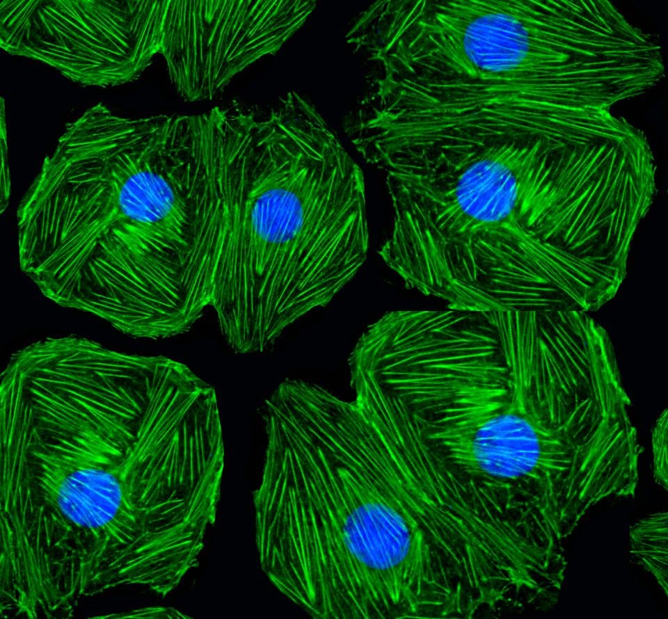
Acti-stain™ 488 phalloidin
Cat. # PHDG1
Acti-stain™ 555 phalloidin
Cat. # PHDH1
Acti-stain™ 670 phalloidin
Cat. # PHDN1
Swiss 3T3 cell stained with anti-vinculin (red), Dapi (blue nucleus) and F-actin is stained with Acti-stain™ 488 (green F-actin, Cat.# PHDG1).
Swiss 3T3 cell with activated Rho, nucleus is stained with Dapi (blue) and F-actin is stained with Acti-stain™ 555 (red F-actin, Cat.# PHDH1).
Mitotic Swiss 3T3 cell, F-actin stained with Acti-stain™ 670 (far-red, Cat. # PHDN1), nuclear DNA stained with Dapi (blue).
Figure 2. Emission and excitation scans for Acti-stain™ phalloidins
Absorbance and fluorescence scan of Acti-stain™ 488. Labeled phalloidin was diluted into methanol and its absorbance and excitation spectra were scanned between 350-750 and 500-750 nm, respectively. Absorbance peaks at 500 nm and fluorescence at 550 nm.
Absorbance and fluorescence scan of Acti-stain™ 555. Labeled phalloidin was diluted into methanol and its absorbance and excitation spectra were scanned between 300-750 and 560-750 nm, respectively. Absorbance peaks at 560 nm and fluorescence at 575 nm.
Absorbance and fluorescence scan of Acti-stain™ 670. Labeled phalloidin was diluted into methanol and its absorbance and excitation spectra were scanned between 300-750 and 600-750 nm, respectively. Absorbance peaks at 625 nm and fluorescence at 675 nm.
Acti-stain phalloidins are the most well characterized phalloidins available. Tabe 1 describes their brightness, photostability, background and affinity constants to F-actin. Compare these performance characteristics to other fluorescent phalloidins and you will see the advantages of using Acti-stain™ for your actin staining requirements.
Table 1. Biochemical characteristics of fluorescent phalloidins
Conjugate | Cat.# | Wavelengths (Ex/Em) | Brightness (AFU*) | Stability to photobleaching(half life in seconds**) | Background (% of total AFU at 100nM**) | Affinity (Kd in nanomolar) |
Fluorescein-phalloidin | na | 485/535 FITC filter set | 432 | 6 | 22 | 72 +/-12 |
Acti-stain™ 488 | PHDG1 | 485/535 FITC filter set | 832 | 57 | 5 | 55 +/-8 |
Acti-stain™ 535 | PHDR1 | 535/585 TRITC filter set | 430 | 27 | 12 | 36 +/-7 |
Acti-stain™ 555 | PHDH1 | 535/585 TRITC filter set | 551 | 46 | 16 | 63 +/-8 |
Acti-stain™ 670 | PHDN1 | 640/680 Cy5 filter set | 837 | 38 | 28 | 12 +/-7 |
* = AFU's measured by quantitative cell imaging. ** = Measured in stained Swiss 3T3 cells in the absence of antifade.
For more information about protocols to fix and stain cells click here.
Manuals and MSDS
Acti-Stain Citations