Anti-Pan Actin Mouse Monoclonal Antibody (Clone 7A8.2.1)

Anti-pan actin: mouse monoclonal
$0.00

Product Uses
This antibody is recommended for detection of actin in a broad spectrum of species such as human, mouse, rat, and bovine.  The following protocols have been tested with this antibody:


 Western BlotImmunocytochemistryELISAImmunoprecipitation
 YesYesNot TestedNot Tested

RRID

AB_2884962

Background and Material

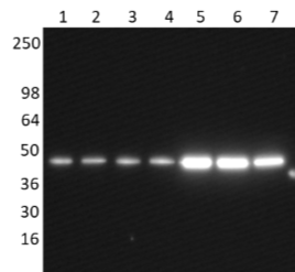
AAN02 is a mouse monoclonal antibody against actin protein. Actin is the major protein of the microfilament cytoskeletal system and is a key protein in various cell motility processes. The immunogen used for antibody production was purified actin protein from rabbit skeletal muscle. A characteristic actin band at 43 kDa is identified on Western blots (Fig. 1).  The antibody has been shown to recognize α-skeletal, α-cardiac, α-smooth muscle, β-cytoplasmic, γ-smooth muscle and  γ- cytoplasmic actin isotypes  (Fig. 1) and has broad species cross-reactivity.  AAN02 is purified by Protein G affinity chromatography and is supplied as a lyophilized white powder. 


Example Results


7A8.2.1_western

Figure 1. Western blot of purified actin and cell extracts probed with anti-actin antibody. Chemiluminescence detection of purified rabbit skeletal muscle actin (10 ng, lane 1), purified bovine cardiac muscle actin (10ng, lane 2), purified chicken gizzard muscle actin (10 ng, lane 3), purified non-muscle human platelet actin (10 ng, lane 4), platelet cell extract (5 µg, lane 5), platelet cell extract (5 µg, lane 6), and A431 cell extract (5 µg, lane 7). The actin band is indicated at 43 kDa. The blot was probed with a 1:1000 dilution of anti-actin antibody. 30 second exposure.
 

7a8.2.1_IF_image

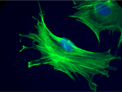
Figure 2. Immunofluorescence images of mouse Swiss 3T3 cells stained with anti-actin antibody. Swiss 3T3 cells were grown to 25% confluency on poly-lysine and laminin coverslips. 3T3 cells were fixed with PFA. Cells were permeabilized with methanol followed by 0.5% Triton X-100 as described in the method. Immunofluorescence staining using 1:500 dilution of anti-actin antibody in PBS is shown (green). The primary antibody was detected with a 1:500 dilution of goat anti-mouse alexfluor-488 conjugated antibody. DNA (blue) was stained with 50 nM DAPI in PBS. Image was taken with a 100X objective lens.
 

7A8.2.1_with_different_fixation_methods_v3.0


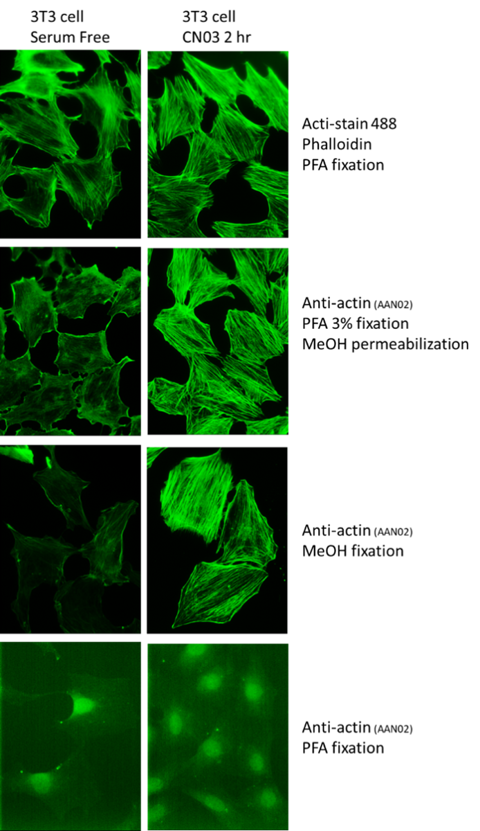
Figure 3. Comparison of fixation strategies used for Immunofluorescence images of mouse Swiss 3T3 cells stained with anti-actin antibody. Swiss 3T3 fibroblasts were plated on glass coverslips, grown to 30% confluency in DMEM plus 10% FBS and serum starved for 24 h in media containing 1% FBS followed by 24 h in serum free media. Cells were treated with a buffer control or 1 µg/ml of the Rho Activator (CN03) for 2 h at 37°C/5% CO2.to induce stress fibers. The 3T3 cells were then fixed with the following methods 1. PFA / phalloidin staining, 2. PFA-MeOH / AAN02 staining, 3. MeOH / AAN02 staining, 4. PFA / AAN02 staining. For condition 2 PFA fixation was followed by permeabilization with methanol as described in the method. Immunofluorescence staining using a 1:500 dilution of anti-actin antibody is shown (green). The primary antibody was detected with a 1:500 dilution of goat anti-mouse alexafluor-488 conjugated antibody. Images were taken with a 40X objective lens.


For product Datasheets, MSDSs, and COAs please click on the PDF links below.


Sample Size Datasheet (Cat. AAN02-S):  

Certificate of Analysis:  available upon request


AuthorTitleJournalYearArticle Link
Chen, Yihe et al.The BK Channel Limits the Pro-Inflammatory Activity of MacrophagesCells2024
Reedy, Jennifer L. et al.Fungal melanin suppresses airway epithelial chemokine secretion through blockade of calcium fluxingNature Communications 2024
Eberhard, Daniel et al.Semaphorin-3A regulates liver sinusoidal endothelial cell porosity and promotes hepatic steatosisNature Cardiovascular Research 2024
Przyklenk, Matthias et al.ANTXR1 deficiency promotes fibroblast senescence: implications for GAPO syndrome as a progeroid disorderScientific Reports 2024
Song, Yang et al.Biphasic regulation of epigenetic state by matrix stiffness during cell reprogrammingScience Advances2024
Politiek, Frouwkje A. et al.Molecular and cellular consequences of mevalonate kinase deficiencyBiochimica et Biophysica Acta (BBA)2024
Müller, Lisa et al.Plakophilin 4 controls the spatio-temporal activity of RhoA at adherens junctions to promote cortical actin ring formation and tissue tensionCellular and Molecular Life Sciences2024
Liu, Xiaomin et al.RABV induces biphasic actin cytoskeletal rearrangement through Rac1 activity modulationJournal of Virology2024
Wang, Kui et al.Vascular endothelial cellular mechanics under hyperglycemia and its role in tissue regenerationRegenerative Biomaterials2024
Sen, Buer et al.Nuclear actin structure regulates chromatin accessibilityNature Communications2024
Baudouin, Lucas et al.Antagonistic actions of PAK1 and NF2/Merlin drive myelin membrane expansion in oligodendrocytesGLIA2024
Gao, Da Peng et al.Chronic Alcohol Exposure Alters the Levels and Assembly of the Actin Cytoskeleton and Microtubules in the Adult Mouse HippocampusJournal of Integrative Neuroscience2024
Kamnev, Anton et al.Coordinated ARP2/3 and glycolytic activities regulate the morphological and functional fitness of human CD8+ T cellsCell reports2024
Yin, Xiufeng et al.CFL1 is Implicated in Chronic Myeloid Leukemia Response during Imatinib TherapyJournal of Cancer2024
Medina-Dols, Aina et al.Role of PATJ in stroke prognosis by modulating endothelial to mesenchymal transition through the Hippo/Notch/PI3K axisCell Death Discovery 2024
Li, Yuan Bao et al.Fimbrin associated with Pmk1 to regulate the actin assembly during Magnaporthe oryzae hyphal growth and infectionStress Biology2024
Wang, Lingling et al.Map-1a regulates Sertoli cell BTB dynamics through the cytoskeletal organization of microtubule and F-actinReproductive Biology and Endocrinology 2024
Yu, Jinhan et al.Inhibiting F-Actin Polymerization Impairs the Internalization of Moraxella catarrhalisMicroorganisms 2024
Kinnart, Inge et al.Elevated α-synuclein levels inhibit mitophagic fluxnpj Parkinson's Disease2024
Omer, Safia et al.Ninein promotes F-actin cup formation and inward phagosome movement during phagocytosis in macrophagesMolecular biology of the cell2024
Gorshkova, Irina N. et al.Human apoA-I[Lys107del] mutation affects lipid surface behavior of apoA-I and its ability to form large nascent HDLJournal of Lipid Research2023
Prasad, Vibhu et al.Enhanced SARS-CoV-2 entry via UPR-dependent AMPK-related kinase NUAK2Molecular cell2023
Rakotomamonjy, Jennifer et al.PCDH12 loss results in premature neuronal differentiation and impeded migration in a cortical organoid modelCell reports2023
Du, William W. et al.Nuclear Actin Polymerization Regulates Cell Epithelial-Mesenchymal TransitionAdvanced Science2023
Levy-Myers, Reuben et al.An independent regulator of global release pathways in astrocytes generates a subtype of extracellular vesicles required for postsynaptic functionScience Advances2023
Faria, Lídia et al.Activation of an actin signaling pathway in pre-malignant mammary epithelial cells by P-cadherin is essential for transformationDisease Models & Mechanisms2023
Romero, Luis O. et al.Linoleic acid improves PIEZO2 dysfunction in a mouse model of Angelman SyndromeNature Communications2023
Lohraseb, Iman et al.Global ubiquitinome profiling identifies NEDD4 as a regulator of Profilin 1 and actin remodelling in neural crest cellsNature Communications2022
Fang, Shi et al.RhoBTB1 reverses established arterial stiffness in angiotensin II–induced hypertension by promoting actin depolymerizationJCI Insight2022
Garcia, Alix et al.miR-204-5p and Platelet Function Regulation: Insight into a Mechanism Mediated by CDC42 and GPIIbIIIaThrombosis and haemostasis2021
Nanamatsu, Azuma et al.Vas******** Induces Urinary Uromodulin Secretion by Activating PKA (Protein Kinase A)Hypertension2021
Wang, Dawei et al.Capping protein regulates endosomal trafficking by controlling f-actin density around endocytic vesicles and recruiting rab5 effectorseLife2021

 If you have any questions concerning this product, please contact our Technical Service department at tservice@cytoskeleton.com