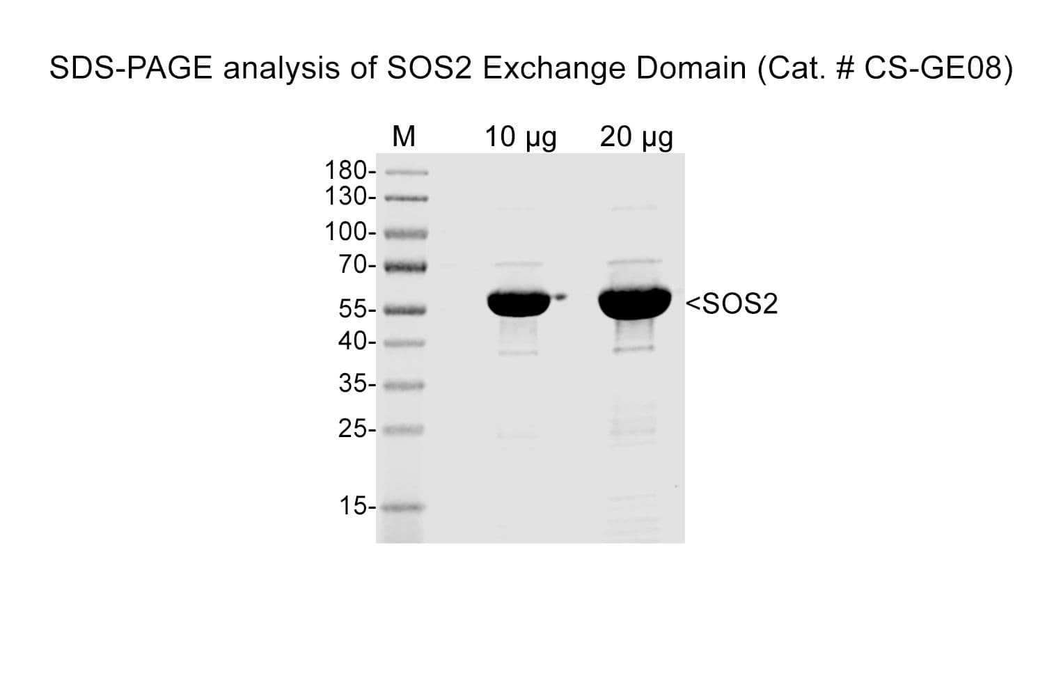
SOS2 exchange domain (563-1051) protein (Human recombinant)

SOS2 exchange domain (563-1051) protein (Human recombinant)
$0.00

* Limited stock available.  If stock is not available, Cytoskeleton will produce a new batch upon request.  Minimum order will apply.  Inquire for more information.


Product Uses

  • Study inhibitors of SOS2 GTP exchange activity
  • Biochemical characterization of SOS2 specific protein interactions
  • Study of SOS2 GEF activity with different GTPases
  • Compare differences in SOS1 and SOS2 exchange activities

Materials

The human SOS2 exchange domain (563-1051) protein (SOS2-ExD) has been produced in a bacterial expression system.  The recombinant protein contains a  6x histidine tag (His-tag) at its amino terminus.  The approximate molecular weight of the SOS2-ExD protein is 61 kDa.  The protein is supplied as a lyophilized white powder.

Storage and Reconstitution

The protein should be reconstituted to 50 mM (3.03 mg/ml) by the addition of 33 µl of nanopure water.  The protein will be in the following buffer; 17 mM Tris pH 7.5, 17 mM NaCl, 0.2 mM MgCl2, 2.3% sucrose, 0.3% dextran.  In order to maintain high biological activity of the protein, it is strongly recommended that the protein solution  should be aliquoted into "experiment sized" aliquots and snap frozen in liquid nitrogen. The protein can be stored at -70°C for 6 months.  Avoid repeated freeze-thaw cycles.  The lyophilized protein is stable at 4°C for 1 year when stored desiccated (<10% humidity).

Purity

Protein purity is determined by scanning densitometry of Coomassie blue stained protein on a 4-20% polyacrylamide gradient gel.  The SOS2-ExD protein was determined to be >90% pure. (see Figure 1).

Figure 1.  SOS2 Protein Purity Determination

Figure 1: Double-helical structure of actin filaments

Legend:  A 10 µg sample of recombinant SOS2-ExD protein (molecular weight approx. 61 kD) was separated by electrophoresis in a 4-20% SDS-PAGE system.  The protein was stained with coomassie blue. Protein quantification was determined using the Precision Red Protein Assay Reagent (Cat.# ADV02).  Mark12 molecular weight markers are from Invitrogen.


Biological Activity Assay

The biological activity of SOS2-ExD can be determined from its ability to catalyze nucleotide exchange on Ras isoforms (N,H,K) using the nucleotide exchange assay with Bodipy-GDP and excess GDP or GTP.  A standard biological assay for monitoring the biological activity of K-Ras4B is an exchange assay utilizing the 2X Exchange Buffer and either the human SOS1 GEF domain (Cat.# CS-GE02) or SOS2 GEF domain (Cat.# CS-GE08)


Reagents

1. Recombinant K-Ras4B protein (Cat.# CS-RS03)

2. Recombinant SOS1-ExD protein (Cat.# CS-GE02)

3. Recombinant SOS2-ExD protein (Cat.# CS-GE08)

4. 2X Exchange Buffer (40 mM Tris pH 7.5, 100 mM NaCl, 20 mM MgCl2 , 0.1 mg/ml BSA, 1.5 µM Bodipy-GTP)

5. Dilution Buffer (20 mM Tris pH 7.5, 50 mM NaCl, 10 mM MgCl2, 0.1 mg/ml BSA)


Equipment

1. Fluorescence spectrophotometer (λex=485 nm, λem=513 nm)

2. Corning 96-well half area plates (Cat. # 3686) or other plate with low protein binding surface.


Method

1. Dilute SOS1-ExD (Cat.# CS-GEO2)  and SOS2-ExD protein (Cat.# CS-GE08) to 1 µM (0.06 mg/ml) with Dilution Buffer.

2. Dilute K-Ras4B (Cat.# CS-RS03) to 50 µM (1.1 mg/ml) with Dilution Buffer.

3. Set up the plate reader for kinetic fluorescence measurements (Excitation wavelength at 485 nm and emission wavelength at 513 nm) with readings every 30 seconds for 30 minutes.

4. Add the following components together and mix well by gentle pipetting


Exchange reaction mix

96 well back plate

2X Exchange Buffer                                 

50µl

dH2O     

26µl

50 µM K-Ras4B 

4 µl

5. Pipette 20 µl of 1 µM SOS1-ExD protein, SOS2-ExD protein, or Dilution Buffer into their respective wells and immediately pipette up and down twice and begin reading the fluorescence.

6. Once the readings are complete and the plate reader file has been saved, the exchange rate can be calculated by reducing the data to Vmax with the software that accompanies the plate reader.


Figure 2.  SOS2-ExD protein mediated Bodipy-GDP exchange on K-Ras4B

Figure 2: Polymerization of actin as measured by pyrene actin fluorescence

Legend: K-Ras4B protein was added to the wells of a 96-well half area plate containing diluted Exchange Buffer and mixed well.  To initiate the exchange reaction, SOS1-ExD protein (blue circles), or SOS2-ExD protein (red squares), was added to the wells, mixed, and fluorescence measurements were obtained using a Molecular Devices M2 Spectramax. The average fluorescence data from triplicate assays were normalized to the fluorescence of buffer at the same time point. The resulting data were plotted as the change in relative fluorescence units (ΔRFU) over time using GraphPad Prism software.

For product Datasheets and MSDSs please click on the PDF links below.

If you have any questions concerning this product, please contact our Technical Service department at tservice@cytoskeleton.com

Coming soon!   If you have any questions concerning this product, please contact our Technical Service department at tservice@cytoskeleton.com