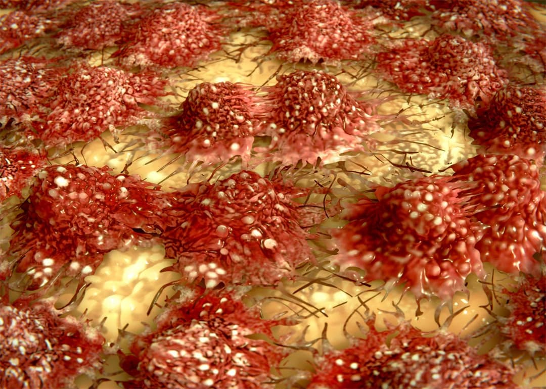
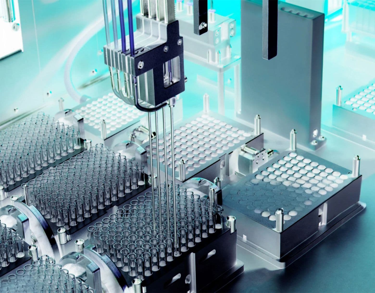
Cytoskeleton, Inc. is a reliable source of compound screening services for kinesin and myosin targeted projects. Over the past decade we have performed numerous contracts for primary and secondary screening, and  SAR based drug development. The basic format is a kinetic ATPase reaction that is used to calculate Vmax data with and without compound. In the kinesin assay microtubules are present to stimulate ATPase, whereas for myosin the S1 domain is generated by proteolytic cleavage and this domain is used either directly on F-actin, or using a reconstituted thin filament / sarcomere assay. Compounds such as blebbistatin, monastrol and omecamtiv mecarbil have been identified with these formats.

Rapidly available target proteins are: Eg5, KHC (KIF5), KIF3C, KIFC3, MKLP1, MKLP2, MCAK, CenPE, Chromokinesin, myosin II (cardiac myosin, smooth myosin, skeletal myosin), non-muscle myosin, and a soluble sarcomere assay which incorporates the Tropomyosin / Troponin complex into a calcium sensitive format. Other assays can also be design and developed and we support all of our services with a dedicated technical services department and years of laboratory experience in the fields of cell biology, cancer biology, cardio-vascular research and neuroscience.

Recently, we have introduced a modular format to the available assays, these are described below, and more details are available by clicking here. An example report can be reviewed by clicking here.

More in depth discussion on myosin and kinesin assays can be found below, or by clicking on the link here.

If you need immediate support, please send an e-mail describing your requirements to tservice@cytoskeleton.com

Examples of completed compound screening contracts: 

1. CenPE and Eg5 kinesin motor IC50 determinations using microtubule stimulated ATPase assays, with monthly analysis of 10 to 30 compounds, results turnaround within one week.

2. Calcium activated sarcomeric myosin S1 ATPase assays with tropomyosin/troponin coated F-actin (thin filaments) as substrate. Determine shift in pCa on addition of 24 compounds at two concentrations, turnaround within 4 weeks.

All projects are recorded in a formal report with Executive summary, Introduction, Methods and Results sections. An example report can be reviewed by clicking here.

For more information click here and then choose ABOUT tab.

Available kinesin and myosin screening modules

Screen name

Amount

Module Code


Kinesin Motor assays (650nm)

96-endpoint (5 IC50 + 1 Control IC50)

CDS050

Kinesin Motor assays (650nm)

96-endpoint (40 duplicates + 4 Controls + 4 blanks)

CDS051

Kinesin Motor assays (360nm)

96-kinetic (5 IC50 + 1 Control IC50)

CDS052

Kinesin Motor assays (360nm)

96-kinetic (40 duplicates + 4 Controls + 4 blanks)

CDS053

Myosin Motor (myosin and F-actin) assays (650nm)

96-endpoint (5 IC50 + 1 Control IC50)

CDS054

Myosin Motor (myosin and F-actin) assays (650nm)

96-endpoint (40 duplicates + 4 Controls + 4 blanks)

CDS055

Myosin Motor (myosin and F-actin) assays (650nm)

96-kinetic (5 IC50 + 1 Control IC50)

CDS056

Myosin Motor (myosin and F-actin) assays (650nm)

96-kinetic (40 duplicates + 4 Controls + 4 blanks)

CDS057

Soluble sarcomere (myosin, tropomyosin/troponin and F-actin) assays (650nm)

96-endpoint (5 IC50 + 1 Control IC50)

CDS058

Soluble sarcomere (myosin, tropomyosin/troponin and F-actin) assays (650nm)

96-endpoint (40 duplicates + 4 Controls + 4 blanks)

CDS059

Soluble sarcomere (myosin, tropomyosin/troponin and F-actin) assays (650nm)

96-kinetic (5 IC50 + 1 Control IC50)

CDS060

Soluble sarcomere (myosin, tropomyosin/troponin and F-actin) assays (650nm)

96-kinetic (40 duplicates + 4 Controls + 4 blanks)

CDS061


More details about myosin and soluble sarcomere assays 

Background

The chemical intervention of myosin has lagged behind other classes of drug targets because of a reluctance to focus on an intracellular molecule which is essential for all human cell types and because in muscle cells it has a high concentration which could potentially reduce the effects of a drug. In the last ten years this situation has changed with the realization that a compound can infiltrate deep into dense muscle tissue and that the myosin family’s diversity allows for quite targeted modulation.

The myosin superfamily is constantly being revised, with one of the latest versions describing 37 classes based on structural domain phylogenies1.  Of these 37 classes, twelve are present in humans and in these twelve classes there are 39 human genes. Of particular significance are the molecular motors involved in cardiac, skeletal and smooth muscle functions which are myosins of Class II; however, there are many more targets that are also of medical value such as myosins of the inner ear (VII), skin (Va) and intestinal microvilli (Ia) (Table I).

Table 1 – Small molecule modulators of myosin

Myosin

Modulator

Mechanism 

Disease relevance 

Ref # 

All Class II isoforms

Blebbistatin

Binds to myosin and inhibits the force cycle at the weakly bound state

not determined

2,3,4

Cardiac II

CK1122534 and Omecamtiv mecarbil

Binds at the cleft between the force generating domain and the ATP binding pocket. Increases the time in the strongly bound state

Heart disease; systolic dysfunction

5,8

Non-muscle II

Ammosamides A&B

Binds irreversibly to an unknown site

not determined

11

Smooth II

CTK2018448

Inhibits myosin and improves renal blood flow

Hypertension

10

Non-muscle Va

Manassantin B

Inhibits the interaction between Myosin Va and melanophillin

Skin coloration disorders

12


Isoform specific myosin drugs

Blebbistatin was one of the earliest identified specific myosin inhibitors and was found to inhibit some class II myosin isoforms with different IC50s2-4. In particular, it has 0.5 to 80 µM affinity for myosin II isoforms4. This report opened up the possibility that compounds could target different isoforms of myosin, and soon after reports emerged that identified several compounds with high affinity for the cardiac myosin II isoform5 and smooth muscle myosin II isoform6. One of these, omecamtiv mecarbil (OM), has been the focus of drug development programs which have reached Phase II clinical trials (clinicaltrials.gov).

It’s important to consider other targets within the sarcomere that interact with myosin and may affect its response to new drugs. In particular, the tropomyosin and troponin C, T, and I complex (T4 complex, Fig. 1) are mediators of the calcium signal for muscle contraction. In concert with the other three members of this complex, Troponin C binds calcium which reduces its affinity for F-actin which allows myosin to bind F-actin and generate force. In an assay format, this has the dramatic effect of increasing the release of phosphate after ATP hydrolysis by up to 100 fold. A selection of these reagents is available from Cytoskeleton on a custom basis (Table 2).

 Figure 1:  Muscle thin filament structure

myosin-newsletter-figure1

Pharmacology

Pharmacologically, myosin is an interesting molecule to study because it has many structural states that constitute the myosin force or duty cycle7 (Fig. 2). In particular, OM has been shown to affect the length of time in the strong force part of the cycle (Fig. 2, green arrow), which is thought to underlie its ability to prolong systolic contraction. Interestingly, diastolic contraction is not affected by OM8. This is rather unexpected but clinically useful because heart failure has a predominance of reduced systolic contraction9. In a similar vein, the smooth muscle inhibitor CTK2018448 (acts in the weakly bound part of the duty cycle; Fig. 2, orange arrow) is reported to increase renal blood flow but not limb blood flow, which is useful for hypertensive patients who have renal insufficiency10. These findings indicate that myosin modulators can have useful and somewhat unpredicted manifestations in whole body systems compared to in vitro assays. 

Figure 2: Actin and Myosin Interactions

myosin-newsletter-figure2

References

  1. Richards T.A. and Cavalier-Smith T. 2005. Myosin domain evolution and the primary divergence of eukaryotes. Nature. 436: 1113-1118.
  2. Cheung A., et al. 2002. A small-molecule inhibitor of skeletal muscle myosin II. Nat. Cell Biol. 4: 83-88.
  3. Straight A.F.,  et al. 2003. Dissecting temporal and spatial control of cytokinesis with a myosin II inhibitor. Science. 299: 1743-1747.
  4. Limouze J., et al. 2004. Specificity of blebbistatin: an inhibitor of myosin II. J. Muscle Res. Cell Motil. 25: 337–341.
  5. Niu C., et al. 2004. The cardiac myosin activator, CK1122534, increases contractility in adult cardiac myocytes but does not affect the transient or depend on second messenger signaling. American Heart Association. November 2004. Poster Presentation.
  6. Qian X., et al. 2009. A direct inhibitor of smooth muscle myosin as a novel therapeutic approach for the treatment of systemic hypertension. American Society of Hypertension 24th Annual Scientific Meeting and Exposition. San Francisco, CA. May 2009. Poster Presentation.
  7. Sweeney L.H. and Houdasse A. 2010. Structural and functional insights into the myosin motor mechanism. Annu. Rev. Biophys. 39: 539–557.
  8. Teerlink J.R., et al. 2011. Dose-dependent augmentation of cardiac systolic function with selective cardiac myosin activator, omecamtiv mecarbil: a first in man study. Lancet. 378: 667-675.
  9. Aronson D. and Krum H. 2012. Novel therapies in acute and chronic heart failure. Pharmacol. Ther. 135: 1-17.
  10. Zhao X., et al. 2011. Inhibition of smooth muscle myosin as a novel therapeutic target for hypertension.  J. Pharmacol. Exp. Ther. 339: 307-312.
  11. Hughes C.C., et al. 2009. Ammosamides A and B target myosin. Angew. Chem. Int. Ed. Engl. 48: 728 –732.
  12. Chang H., et al 2012. Manassantin B inhibits melanosome transport in melanocytes by disrupting the melanophilin-myosin Va interaction. Pigment Cell Melanoma Res. 25: 765-772.


More details about Kinesin / microtubule assays

Eukaryotic kinesin motor proteins orchestrate a wide range of kinetic events within a cell.  They have been shown to move cargoes, such as chromosomes and vesicles, along microtubule tracks (1).  They also play a major role in the organization of cytoskeletal architecture as evidenced in the establishment of the microtubule spindle during mitosis (2). 

Kinesins operate by utilizing the energy of ATP hydrolysis to move along their microtubule (MT) substrates.  Once a kinesin motor binds to its MT track, the ATPase rate of the motor is often enhanced several hundred to several thousand fold (3).   MT activated kinesin ATPase is a major parameter in  motor function and serves as a powerful method to monitor / study kinesin activity under various experimental conditions.

As part of it’s Cytoskeleton Motor Werks (CMW) line of research reagents, Cytoskeleton Inc. has developed two assays to measure microtubule stimulated kinesin and dynein ATPase. First, the kinesin end-point assay is an extremely quick and economical way to measure inorganic phosphate (Pi) generated during the microtubule activated ATPase activity of kinesin motor proteins.  Large numbers of assays can be performed simultaneously in a homogenous reaction, making the assay highly suitable for HTS applications.  The assay is an adaptation of the method of Kodama, T. et al. J. Biochem. 99: 1465-1472 (1986). Essentially liberated phosphate is detected by a malachite green / molybdate complex which turns a green color on binding to phosphate.

The  kinetic kinesin ATPase assay is based on the Kinesin ELIPA (Enzyme Linked Inorganic Phosphate Assay) Biochem Kit offered by Cytoskeleton Inc.  The assay is an adaptation of a method originally described by Webb for the measurement of glycerol kinase plus D-glyceraldehyde ATPase activity and for actin activated myosin ATPase (4).  The assay is based upon an absorbance shift (330 - 360 nm) that occurs when 2-amino-6-mercapto-7-methylpurine ribonucleoside (MESG) is catalytically converted to 2-amino-6-mercapto-7-methyl purine in the presence of inorganic phosphate (Pi).  The reaction is catalysed by purine nucleoside phosphorylase (PNP).  One molecule of inorganic phosphate will yield one molecule of 2-amino-6-mercapto-7-methyl purine in an essentially irreversible reaction (5).  Thus, the absorbance at 360 nm is directly proportional to the amount of Pi generated in the kinesin ATPase reaction.  Figure 3 shows a typical set of results from a kinesin heavy chain motor domain protein MT activated ATPase ELIPA.

Figure 3 - Kinesin Heavy Chain ELIPA

Kinesin-Heavy-Chain-ELIPA

Method:  The reactions were  conducted in a 96 well plate format (300 ul reaction volumes).  Each reaction contains 94 nM kinesin heavy chain motor domain protein (cat # KR01), 0.66 uM taxol stabilized microtubules (cat # MT002), 0.2 mM MESG, 0.3 U PNP, 15 µM taxol, 15 mM PIPES pH 7, 5 mM MgCl2, 0.6 mM ATP.  Control reactions were carried out in the absence of motor protein and in the absence of MTs, these reactions gave readings of <0.02 (data not shown). Reactions were measured in a SpectraMax 250 (Molecular Devices) set in kinetic mode and 360 nm absorbance wavelength. Readings were taken at room temperature once every 30 seconds for a total reaction time of 20 minutes.


References

1)      Vale, R.D. & Fletterick, R.J. 1997.  The Design Plan of Kinesin Motors. Ann. Rev. Cell Dev. Biol. 13: 745-777.

2)      Endow, S. A. 1999.  Microtubule Motors in Spindle and Chromosome Motility.  Eur. J. Biochem 262: 12-18.

3)      Johnson, K.A. & Gilbert, S.P. 1995.  Pathway of the Microtubule-Kinesin ATPase.  Biophysical Journal 68: 173s - 179s.

4)      Webb, M.R. 1992. A continuous spectrophotometric assay for inorganic phosphate and for measuring phosphate release kinetics in biological systems.  Proc. Natl. Acad. Sci. USA 89: 4884-4887.

5)      Analytical Biochemistry 1997 246: 86-95

6)      Lockhart, A & Cross, R.A. 1996.  Kinetics and Motility of the Eg5 Microtubule Motor. Biochemistry 35: 2365 - 2373.