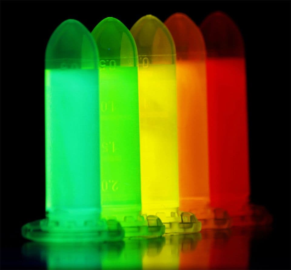
Fluorescent & labeled actins
Cyto offers a premier selection of biotinylated and fluorescent actin proteins designed to meet the exacting standards of leading cell biologists and biophysicists. Whether you're pushing the boundaries of fundamental research or building the next breakthrough in synthetic biology, our actin tools give you the precision and flexibility you demand.
Use them to:
- Recreate in vitro bio-membranes with biologically relevant accuracy
- Dissect the molecular mechanics of cytoskeletal force generation
- Model the dynamic architecture of the cell cortex
- Track free barbed ends to illuminate in vivo actin polymerization
- Probe actin-binding protein interactions with clarity and control
- Power next-gen applications in nanotechnology and bioengineering
- Follow in vitro actin polymerization under varied experimental conditions
When precision, performance, and publication-ready results matter— scientists choose Cyto.
