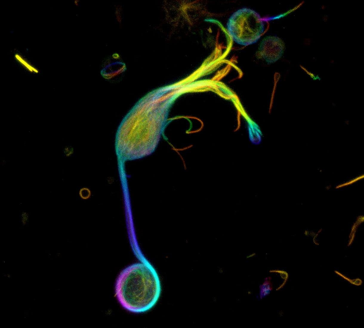
Spirochrome's New SPY™ Probes

In 2015, Cytoskeleton introduced multiple live cell imaging probes from Spirochrome Ltd. These revolutionary probes simplifed and stream-lined the live cell imaging of microtubules (SiR-Tubulin), F-actin (SiR-Actin), lysosomes (SiR-Lysosome), and chromosomal DNA (SiR-DNA).  Based on the proprietary fluorophore silicon rhodamine (SiR), the original SiR probes introduced a new era in live cell imaging of cytoskeletal proteins, lysosomes, and DNA. These fluorogenic, bright, and photostable probes with low background and cytotoxicity represented a major improvement in live cell imaging probes that had been use for decades. These game-changing SiR probes are defined by multiple positive features and improvements over other live cell imaging probes, including:


  1. Cell Permeability (No Transfections)
  2. Photostability
  3. Very Low Background
  4. Super-resolution Compatibility (STED, SIM)
  5. Far-Red Fluorophore (Cy5-compatible)
  6. Fluorogenic Nature
  7. No Cytotoxicity

Now in 2019, Cytoskeleton is excited to introduce the next generation of Spirochrome’s live cell imaging probes – the SPY™ probes. SPY™ probes improve upon the SiR and SiR700 live cell imaging probes in the far-red channels while also expanding the fluorophore labeing options for the study of F-actin, microtubules, and DNA in living cells. Fluorogenic live cell fluorescent probes are now available in the green, orange, red and far-red channels  (see excitation and emission spectra figures below).

93X STED-imaged HeLa cell labeled with SPY505-DNA (green) and SPY555-actin (magenta). Image provided courtesy of Spirochrome.


SPY™ probes capitalize on all of the benefits and advantages of SiR probes, while also offering substantial improvements which include:


  1. Fluorogenic Nature – background is even less than that of SiR and SIR700 probes
  2. Multiple Fluorophore Options (e.g., far-red, orange, red, and green)
  3. Sensitivity and Selectivity
  4. Improved Cell Permeability
  5. Follows the same protocols as for SiR with shorter incubation times and lower concentrations (probes last longer and are less likely to affect cytoskeletal dynamics)
  6. No Cytotoxicity

HeLa Cell stained with SPY555-Actin (magenta), SPY595-DNA and SPY650-Tubulin

New SPY Actin Probes

SPY555-actin (Cat. #CY-SC202) is an F-actin probe with performance similar to SiR-actin (Cat. # CY-SC001) but in the tetramethylrhodamine (TMR) channel with a recommended optimal excitation setting of 555 nm and emission setting of 580 nm. SPY555-actin should be imaged in a standard TMR or Cy3 channel using the same staining protocol as for SiR-actin (Cat. # CY-SC001). The recommended live cell staining conditions from Spirochrome scientists are 100 nM for 2 hours.

HeLa cell stained with SPY555-actin. Image courtesy of Spirochrome

New SPY Tubulin Probes

SPY555-tubulin (Cat. #CY-SC203) is a microtubule probe that has performance superior to SiR-tubulin (Cat.# CY-SC007) but in the tetramethylrhodamine (TMR) channel with a recommended optimal excitation setting of 555 nm and emission setting of 580 nm. SPY555-tubulin should be imaged in a standard TMR or Cy3 channel using the same general staining protocol as for SiR-tubulin (Cat. # CY-SC002). However, as SPY555-tubulin is much more cell permeable than SiR-tubulin, the recommended live cell staining conditions from Spirochrome scientists are 10 nM for 2 hours.

HeLa cell stained with SPY555-tubulin. Image courtesy of Spirochrome

SPY650-tubulin (Cat. #CY-SC503) is a modified version of SiR-tubulin (Cat. # CY-SC002) as it is also conjugated to a SiR-fluorophore with an optimal excitation setting of 650 nm and an emission setting of 675 nm. SPY650-tubulin stains microtubules much better than SiR-tubulin (about 10 fold) at equivalent concentrations, and it has a lower background fluorescence signal. SPY650-tubulin should be imaged in a standard Cy5 channel using the same staining protocol as for SiR-tubulin (Cat. # CY-SC002). Due to its improved cell permeability over SiR-tubulin (Cat. # CY-SC002), SPY650-tubulin’s recommended live cell staining conditions from Spirochrome scientists are 10 nM for 2 hours.

New SPY DNA Probes

SPY505-DNA (Cat. #CY-SC101) is a green channel DNA probe with an optimal excitation setting of 510 nm and emission setting of 530 nm. SPY505-DNA should be imaged in a fluorescein isothiocyanate (FITC) or yellow fluorescent protein (YFP) channel using the same staining protocol as used for SiR-DNA (Cat. # CY-SC007). The recommended live cell staining conditions from Spirochrome scientists are 100 nM for 2 hours.

HeLa cell stained with SPY505-DNA. Image courtesy of Spirochrome.

SPY555-DNA (Cat. #CY-SC201) is a trimethylrhodamine (TMR) channel DNA probe with an optimal excitation setting of 555 nm and emission setting of 580 nm. SPY555-DNA should be imaged in a TMR or Cy3 channel using the same staining protocol as used for SiR-DNA (Cat. # CY-SC007). The recommended live cell staining conditions from Spirochrome scientists are 100 nM for 2 hours.

HeLa cell stained with SPY555-DNA. Image courtesy of Spirochrome

SPY595-DNA (Cat. #CY-SC301) is an "Alexa 594" channel DNA probe with an optimal excitation setting of 605 nm and emission setting of 645 nm. SPY595-DNA should be imaged in an “Alexa594”-compatible or TexasRed channel using  the same staining protocol as used for SiR-DNA (Cat. # CY-SC007). The recommended live cell staining conditions from Spirochrome scientists are 100 nM for 2 hours.

SPY650-DNA (Cat. #CY-SC501) is a modified version of SiR-DNA (Cat. # CY-SC007). SPY650-DNA contains a SiR-fluorophore analogue with an optimal excitation setting of 650 nm and emission setting of 675 nm. At equivalent concentrations, SPY650-DNA stains cell nuclei much better than SiR-DNA (Cat. # CY-SC007) (3-6 fold brighter without increased toxicity). SPY650-DNA should be imaged in a standard Cy5 channel using the same staining protocol as used for SiR-DNA (Cat. # CY-SC007). The recommended live cell staining conditions from Spirochrome scientists are 100 nM for 2 hours.

SPY700-DNA (Cat. #CY-SC601) is a modified version of SiR700-DNA (Cat. # CY-SC015). SPY700-DNA contains a SiR700-fluorophore with an optimal excitation setting of 690 nm and emission setting of 720 nm. At equivalent concentrations, SPY700-DNA stains cell nuclei much better than SiR700-DNA (Cat. # CY-SC015) (3-6 fold brighter without increased toxicity). SPY700-DNA can be imaged in a standard Cy5 channel, but a further far-red channel yields a much better signal. The same staining protocol as used for SiR700-DNA (Cat. # CY-SC015) should be used for SPY700-DNA. The recommended live cell staining conditions from Spirochrome scientists are 100 nM for 2 hours.

How do SPY™ Probes match up against the competition?

For further information on the subject, please refer to this Nature Chemistry  article   

Article Link Here