This fluorescent phalloidin-based actin staining kit enables precise visualization of F-actin in fixed cells. It includes all necessary fixation buffers for consistent, high-specificity results.
Key characteristics
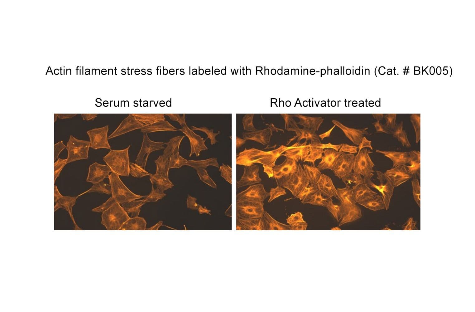
- Bright, specific labeling: Phalloidin probe (Ex: 535 nm, Em: 585 nm) selectively binds polymerized actin, minimizing background noise
- Cross-isotype and species compatibility: Binding unaffected by actin isoforms or species differences
- Streamlined workflow: Standardized staining protocol reduces variability across experiments
- Long-lasting signal: Anti-fade mounting medium preserves fluorescence for imaging