1. Home
  2. About Acti-stain

About Acti-stain™
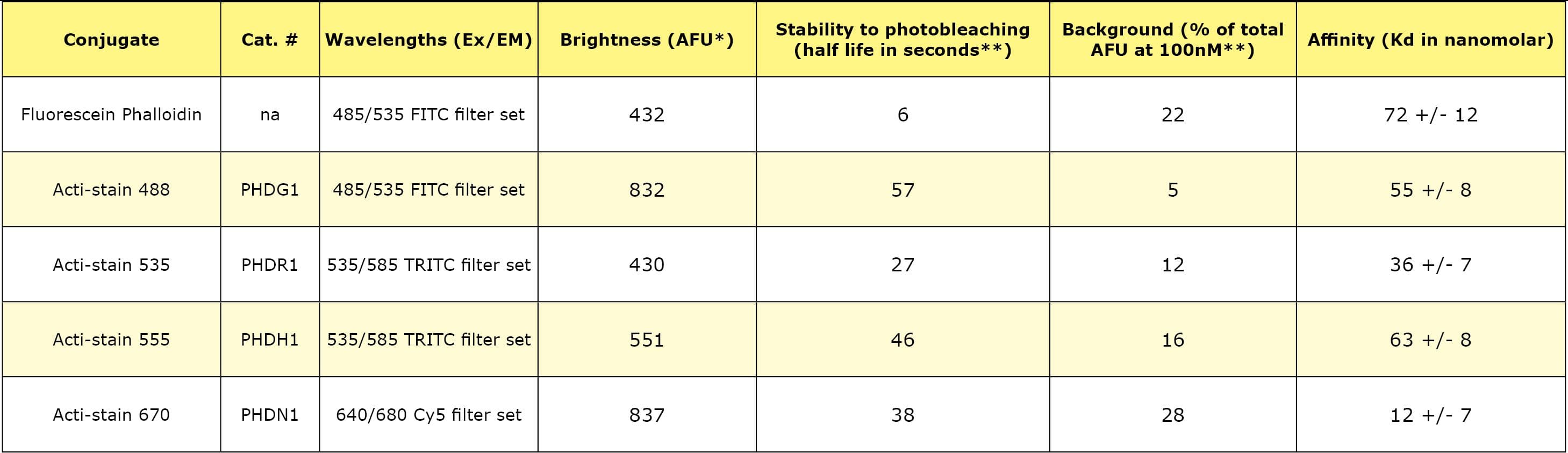
The Acti-stain™ line of fluorescent phalloidins has been developed with an emphasis on creating exceptionally bright and stable probes at an economical price. Side-by-side comparisons with leading competing products demonstrate that you will enjoy savings while not sacrificing the quality of the stain when switching to an Acti-stain™ probe. Additionally, these probes are compatible with popular filter sets such as FITC, TRITC and Cy5. The combination of in-house manufacturing, stringent quality control and convenient packaging provides a great value. Give them a try and see for yourself.

Product Uses Include

  • Stain F-actin in fixed cells
  • Stabilize actin filaments in vitro
  • Visualize actin filaments in vitro

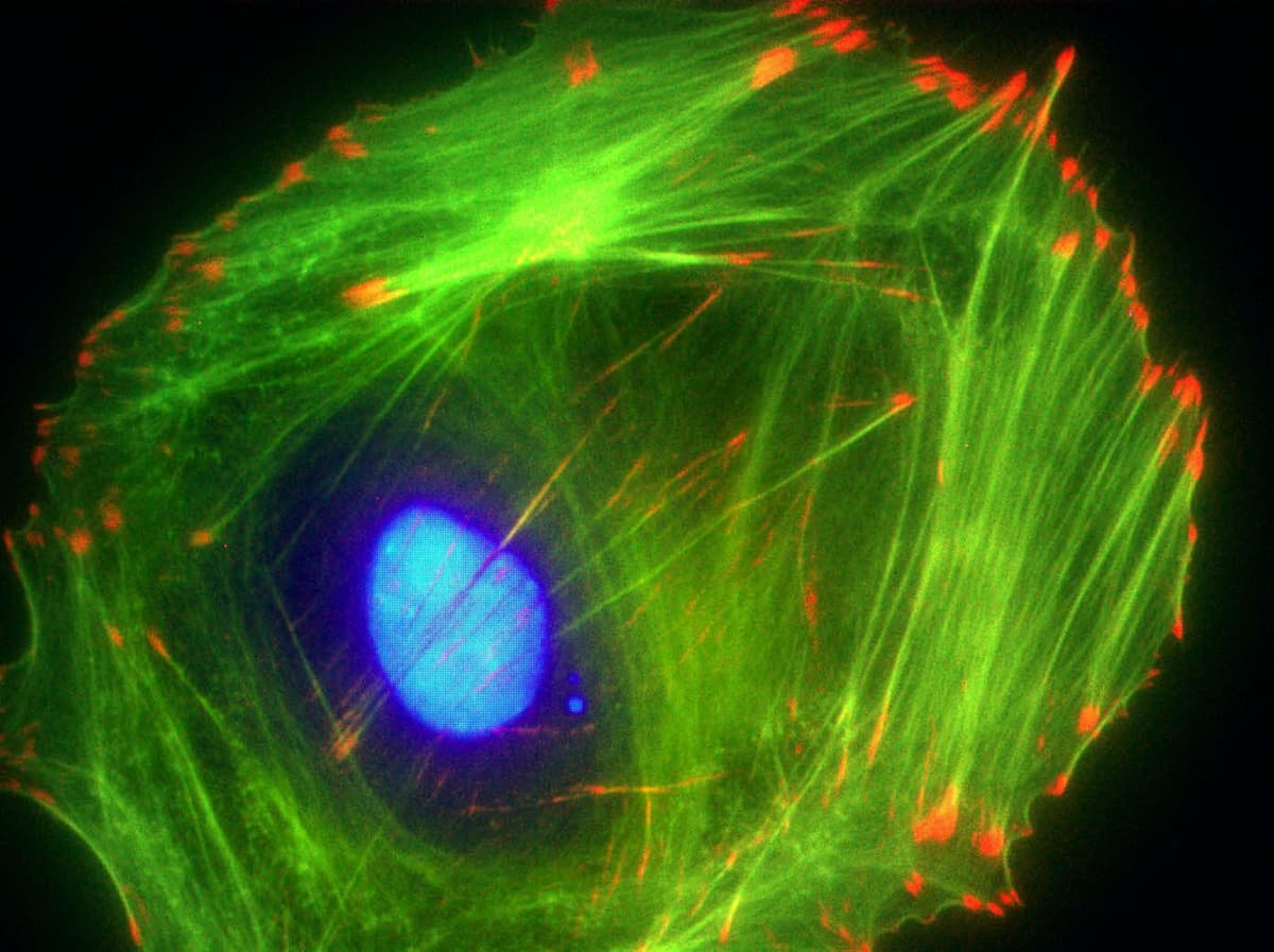
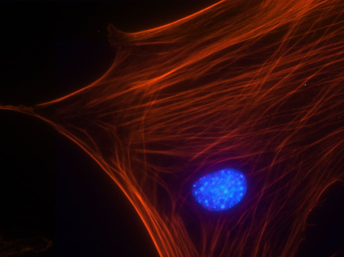
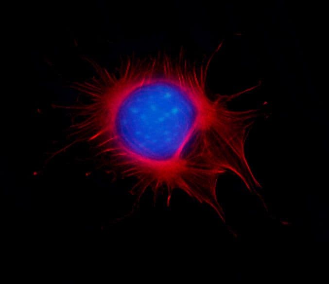
Actin staining is very useful in determining the structure and function of the cytoskeleton in living and fixed cells. The actin cytoskeleton is a very dynamic and labile structure in the living cell, but it can be fixed with paraformaldehyde prior to probing or staining for actin structures.

Acti-stain™ 488 phalloidin
Cat. # PHDG1

Sanity Image
Swiss 3T3 cell stained with anti-vinculin (red), Dapi (blue nucleus) and F-actin is stained with Acti-stain™ 488 (green F-actin, Cat.# PHDG1).

Specifications:

Absorbance and fluorescence scan of Acti-stain™ 488. Labeled phalloidin was diluted into methanol and its absorbance and excitation spectra were scanned between 350-750 and 500-750 nm, respectively. Absorbance peaks at 500 nm and fluorescence at 550 nm.

Sanity Image

Acti-stain™ 555 phalloidin
Cat. # PHDH1

Sanity Image
Swiss 3T3 cell with activated Rho, nucleus is stained with Dapi (blue) and F-actin is stained with Acti-stain™ 555 (red F-actin, Cat.# PHDH1).

Absorbance and fluorescence scan of Acti-stain™ 555. Labeled phalloidin was diluted into methanol and its absorbance and excitation spectra were scanned between 300-750 and 560-750 nm, respectively. Absorbance peaks at 560 nm and fluorescence at 575 nm.

Sanity Image

Acti-stain™ 670 phalloidin
Cat. # PHDN1

Sanity Image
Mitotic Swiss 3T3 cell, F-actin stained with Acti-stain™ 670 (far-red, Cat. # PHDN1), nuclear DNA stained with Dapi (blue).

Absorbance and fluorescence scan of Acti-stain™ 670. Labeled phalloidin was diluted into methanol and its absorbance and excitation spectra were scanned between 300-750 and 600-750 nm, respectively. Absorbance peaks at 625 nm and fluorescence at 675 nm.

Sanity Image

Acti-stain phalloidins are the most well characterized phalloidins available. Tabe 1 describes their brightness, photostability, background and affinity constants to F-actin. Compare these performance characteristics to other fluorescent phalloidins and you will see the advantages of using Acti-stain™ for your actin staining requirements.

Table 1. Biochemical characteristics of fluorescent phalloidins

* = AFU's measured by quantitative cell imaging. ** = Measured in stained Swiss 3T3 cells in the absence of antifade.

For more information about protocols to fix and stain cells click here.

Manuals and MSDS

To view the Manuals and MSDS sheets of each product, please click on the links below:

Acti-stain 488 phalloidin (Cat # PHDG1)

Acti-stain 555 phalloidin (Cat # PHDH1)

Acti-stain 670 phalloidin (Cat # PHDN1)

Acti-Stain Citations

Takaya K, Okabe K, Sakai S, Aramaki-Hattori N, Asou T, Kishi K. 2024. Salicylate induces epithelial actin reorganization via activation of the AMP-activated protein kinase and promotes wound healing and contraction in mice. Scientific Reports. 14(1). doi:https://doi.org/10.1038/s41598-024-67266-5.

Wang C, Zhao X, Zhao L, Wang Y, Jia Y, Zhang X, Ma W. 2023. PKCζ phosphorylates VASP to mediate chemotaxis in breast cancer cells. Experimental Cell Research. 433(2):113823. doi:https://doi.org/10.1016/j.yexcr.2023.113823.

Phillips AT, Boumil EF, Venkatesan A, Tilstra-Smith C, Bernstein AM. 2023. The formin DAAM1 regulates the deubiquitinase activity of USP10 and integrin homeostasis. European Journal of Cell Biology. 102(Chapter 10):151347. doi:https://doi.org/10.1016/j.ejcb.2023.151347.

Boonrod K, Kuaguim L, Braun M, Müller-Renno C, Ziegler C, Krczal G. 2023. Identification of the Actin-Binding Region and Binding to Host Plant Apple Actin of Immunodominant Transmembrane Protein of “Candidatus Phytoplasma mali.” International Journal of Molecular Sciences. 24(2):968. doi:https://doi.org/10.3390/ijms24020968.

Chang Y, Kong K, Tong Z, Qiao H, Hu Y, Xia R, Zhang J, Zhai Z, Li H. 2023. Aspirin prevents estrogen deficiency-induced bone loss by inhibiting osteoclastogenesis and promoting osteogenesis. Journal of Orthopaedic Surgery and Research. 18(1). doi:https://doi.org/10.1186/s13018-023-03710-y.

Bhosle VK, Sun C, Patel S, Ho TWW, Westman J, Ammendolia DA, Langari FM, Fine N, Toepfner N, Li Z, et al. 2023. The chemorepellent, SLIT2, bolsters innate immunity against Staphylococcus aureus. eLife. 12. doi:https://doi.org/10.7554/elife.87392.

Karvas RM, Zemke JE, Ali SS, Upton E, Sane E, Fischer LA, Dong C, Park K-M, Wang F, Park K, et al. 2023. 3D-cultured blastoids model human embryogenesis from pre-implantation to early gastrulation stages. Cell Stem Cell. 30(9):1148-1165.e7. doi:https://doi.org/10.1016/j.stem.2023.08.005.

Park J, Kravchuk P, Krishnaprasad A, Roy T, Kang EH. 2022. Graphene Enhances Actin Filament Assembly Kinetics and Modulates NIH-3T3 Fibroblast Cell Spreading. International Journal of Molecular Sciences. 23(1):509. doi:https://doi.org/10.3390/ijms23010509.

Cytoskeleton, Inc
Contact UsMy Account

Tel: 303-322-2254

  • Blogs
  • FAQ's
  • Datasheets and MSDS
  • Protocols
  • Learning Documents
  • Videos
  • About Us
  • Careers
  • Distributors
  • All Services
  • Compound Screening
  • Protein Purification
  • Assay Development
  • Post-Translational Modification (PTM) Tools
  • Motor Protein Products
  • Live Cell Imaging Reagents
  • Actin Products
  • ECM Proteins
  • Small G-protein Products
  • Tubulin & FtsZ Products
  • General Protein Tools
Linkedin

© 2026 Cytoskeleton, Inc All Rights Reserved.

Privacy Policy

TEL: 303-322-2254

Log in to your Account
Cytoskeleton, IncCytoskeleton, Inc
Your quote is empty.
My Cart
( Items)
  • Cytoskeleton, Inc
  • Products
    • Featured New Product
    • Small G-protein Products
      • Small G-protein Activators & Inhibitors
      • Small G-protein Activation Assays
      • Small GTPase Affinity Beads
      • Small G-protein GEF/GAP and ELISA Kits
      • Small G-proteins
      • Modulator Proteins
      • Small G-protein Antibodies
      • Small G-protein Buffers
    • Post-Translational Modification (PTM) Products
      • Ubiquitination Tools
      • Acetyl-Lysine Tools
      • SUMOylation Tools
      • Phosphotyrosine Tools
      • Actin Oxidation Tools
      • Antibodies: PTMtrue™
      • Cell Lysis Tools
    • Tubulin & FtsZ Products
      • Tubulin Kits
      • Microtubules & Tubulin Proteins
      • Fluorescent and Labeled Tubulins
      • Microtubule-Associated Proteins (MAPs)
      • Live Cell Tubulin Reagents
      • Tubulin Antibodies
      • FtsZ Proteins
      • Tubulin Drugs and Buffers
    • Actin Products
      • Acti-stain™ Fluorescent Phalloidins
      • Actin Assays & Kits
      • Actin Proteins
      • Fluorescent and Labeled Actin
      • Actin Binding Proteins
      • Live Cell Actin Reagents
      • Actin Oxidation Tools
      • Actin Antibodies
      • Actin Drugs & Buffers
    • Live Cell Imaging Reagents
      • Live Cell Actin Reagents
      • Live Cell Tubulin Reagents
      • Small G-protein Activators & Inhibitors
      • Nuclear DNA Probes
      • Live Cell Membrane Probes
        • Plasma Membrane Probes
        • Membrane Tension Probes
        • Membrane Polarity Probes
      • Live Cell Mitochondrial Reagents
      • ECM Proteins
        • Fluorescent Fibronectin
        • Fluorescent Laminin
      • Live Cell Lipid Droplets
      • Other Organelles
      • Dyes/Substrates
    • ECM Proteins
      • Fluorescent Fibronectin
      • Fluorescent Laminin
    • Motor Protein Products
      • Motor Kits and Assays
      • Kinesin Proteins
      • Myosin Proteins
      • Dynein Proteins
      • Motor Protein Drugs & Buffers
    • General Protein Tools
      • Protein Quantitation
      • Cell Lysis Tools
      • ATPase & GTPase Assays
      • Other Protein Tools
    • GO-Blot V2 - Fully Automated And Programmable Western Blot Processor
  • Custom Services
    • All Services
    • Compound Screening
      • Tubulin Polymerization Assay Services
      • Kinesin ATPase Screening Services
      • Myosin ATPase Screening Services
      • GTP Exchange Factor (GEF) Assay Services
      • GTPase Activating Protein (GAP) Assay Services
      • FtsZ GTPase and Polymerization Assay Services
      • GTPase Activation Screening Services
    • Protein Purification
    • Assay Development
  • Resources
    • Resource Hub
      • Blogs
      • Videos
      • Protocols
      • Learning Documents
        • Ebook
    • FAQs
    • Datasheets and MSDS
  • About Us
    • About Us
    • Careers
    • Distributors
  • Contact Us
Your cart is empty.