As part of the Signal-Seeker™ product line, phosphotyrosine affinity beads have been optimized to detect endogenous levels of phosphotyrosine modified proteins, which often represent <1% of the target protein. Phosphotyrosine affinity beads comprise an anti-phosphotyrosine antibody (Clone 27B10) that has been chemically conjugated to Protein G beads. Validation studies have shown that these beads can immunoprecipitate a wide range of phosphotyrosine proteins without detectable leaching of either heavy or light chains in an IP assay. A comprehensive Signal-Seeker™ Phosphotyrosine Detection Kit is also available (BK160) and is recommended for first time users.
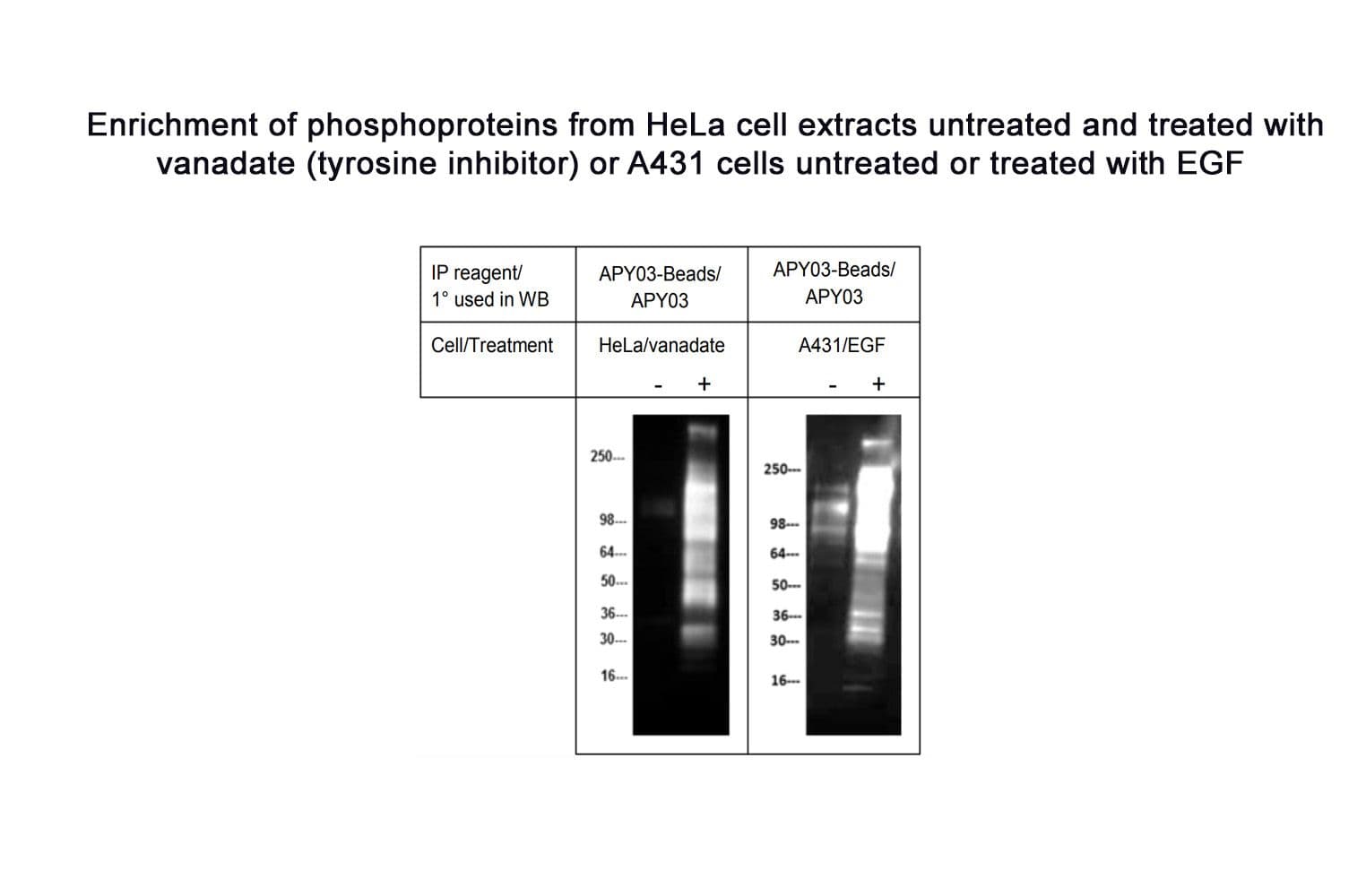
Enrichment of phosphotyrosine proteins from HeLa and A431 cell lysates
HeLa cells were either treated or untreated with 100 μM of H2O2 activated sodium othovanadate for 30 minutes. A431 cells were serum starved for 24 hours before treatment with EGF (50 ng/ml for 5 min). Cell lysate was obtained using RIPA buffer. 1 mg of cell lysate was incubated with 30 µl of APY03-Bead slurry. Eluted proteins were resolved by SDS-PAGE and transferred to PVDF membrane. Anti-phosphotyrosine antibody APY03 (1:500) and goat-anti-mouse secondary (1:20000, Jackson Labs # 115-035-068) were used to detect tyrosine phosphorylated proteins. Western blot was developed with SuperSignal West Dura chemiluminescent reagent (Thermo Scientific) with an exposure time of 20 seconds.
To see the full Western blot protocol, see the product datasheet.
Figure 1: Enrichment of Phosphotyrosine Proteins from Cell Lysates
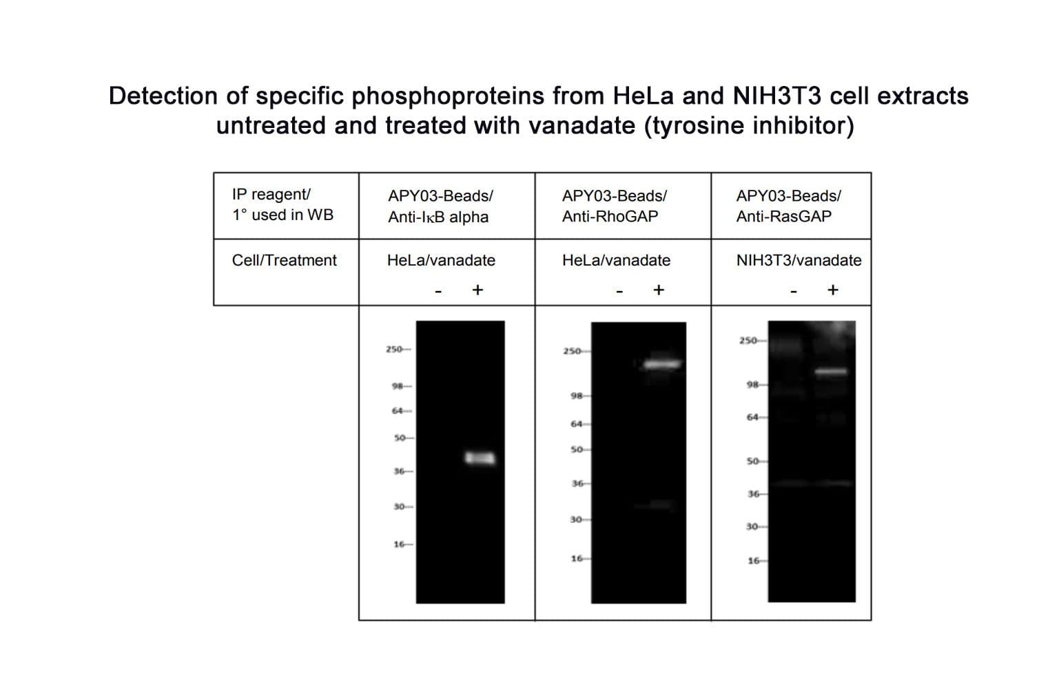
Enrichment of phosphotyrosine modified IκB alpha, RhoGAP and RasGAP proteins from HeLa and NIH3T3 cell lysates
Hela cells were either treated or untreated with 100 µM of H2O2 activated sodium orthovanadate for 30 minutes. NIH3T3 cells were either treated or untreated with 100 μM of H2O2 activated sodium othovanadate for 10 minutes . Cell lysate was obtained using RIPA buffer. 1 mg of cell lysate was incubated with 30 µl of APY03-Bead slurry. Eluted proteins were resolved by SDS-PAGE and transferred to PVDF membrane. Primary antibodies, anti-IkB alpha (1:1000, BD Biosciences # 610690), anti-RhoGap (1:1000, Millipore # 05-378) and anti-RasGap (1:1000, BD Biosciences # 610040) and goat-anti-mouse secondary (1:20000, Jackson Labs # 115-035-068) were used in western blot analysis. Western signal was developed with SuperSignal West Dura chemiluminescent reagent (Thermo Scientific) and exposure time for IkB alpha, RhoGAP and RasGAP were 5, 10 and 3 minutes respectively. Figure 2 shows that APY03-Beads are able to immunoprecipitate endogenous tyrosine phosphorylated IkB alpha (~38 kDa) and RhoGAP (~190 kDa) from Hela cells treated with sodium orthovanadate and tyrosine phosphorylated RasGAP (~120 kDa) from NIH3T3 treated with sodium orthovanadate.
Figure 2: Enrichment of Phosphotyrosine Modified IkB alpha, RhoGAP and RasGAP from Cell Lysates.
Amount:
Each package contains enough phosphotyrosine beads for 40 reactions.
For more information contact: signalseeker@cytoskeleton.com
Associated Products:
Signal-Seeker™ Phosphotyrosine Detection Kit (Cat. # BK160)
Signal-Seeker™ Ubiquitination Detection Kit (Cat. # BK161)
Signal-Seeker™ SUMOylation 2/3 Detection Kit (Cat. # BK162)
Signal-Seeker™: BlastR™ Rapid Lysate Prep Kit (Cat. # BLR01)
Signal-Seeker™: PTMtrue™ Phosphotyrosine Antibody (Cat.# APY03)
Visit our Signal-Seeker™ Tech Tips and FAQs page for technical tips and frequently asked questions regarding this and other Signal-Seeker™ products click here
If you have any questions concerning this product, please contact our Technical Service department at tservice@cytoskeleton.com