Rac1 Pull-down Activation Assay Biochem Kit (bead pull-down format) - 50 Assays

Rac1 Activation Assay Biochem Kit (bead pull-down format) - 50 Assays
$0.00

Product Uses Include

  • Analysis of in vivo Rac1 activation levels.
  • Detection of compounds and proteins that enhance Rac1 activity
  • Detection of compounds and proteins that inhibit Rac1 activity

Introduction
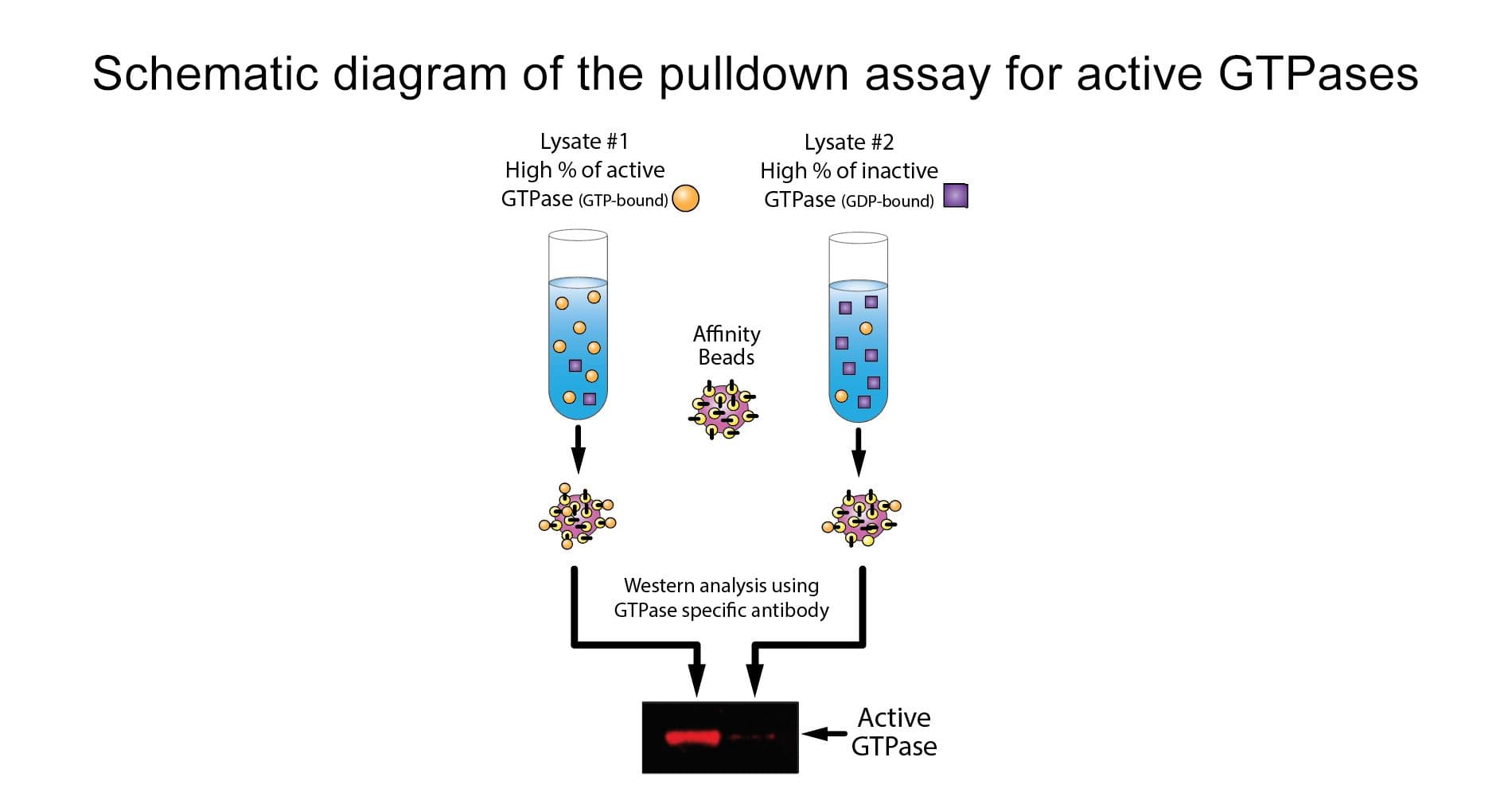
The Rho switch operates by alternating between an active, GTP-bound state and an inactive, GDP-bound state.  Understanding the mechanisms that regulate activation / inactivation of the GTPases is of obvious biological significance and is a subject of intense investigation.  The fact that many Rho family effector proteins will specifically recognize the GTP bound form of the protein has been exploited experimentally to develop a powerful affinity purification assay that monitors Rac and Cdc42 protein activation.  The assay uses the Cdc42/Rac Interactive Binding (CRIB) region (also called the p21 Binding Domain, PBD) of the Cdc42 / Rac effector protein, p21 activated kinase 1 (PAK).  The CRIB/PBD protein motif has been shown to bind specifically to the GTP-bound form of Rac and/or Cdc42 proteins.  The fact that the PBD region of PAK has a high affinity for both GTP-Rac and GTP-Cdc42 and that PAK binding results in a significantly reduced intrinsic and catalytic rate of hydrolysis of both Rac and Cdc42 make it an ideal tool for affinity purification of GTP-Rac and GTP-Cdc42 from cell lysates.  The PAK-PBD protein supplied in this kit corresponds to residues 67-150.  This includes the highly conserved CRIB region (aa 74-88) plus sequences required for the high affinity interaction with GTP-Rac and GTP-Cdc42.  The PAK-PBD is in the form of a GST fusion protein, which allows one to "pull-down" the PAK-PBD/GTP-Rac (or GTP-Cdc42) complex with glutathione affinity beads.  The assay therefore provides a simple means of quantitating Rac or Cdc42 activation in cells.  The amount of activated Rac is determined by a Western blot using a Rac-specific antibody.


Kit contents
The kit contains sufficient materials for 50 assays, depending on assay setup, and includes reagents for positive and negative controls. The following components are included:

  1. GST-tagged PAK-PBD protein on colored agarose beads (Cat. # PAK02)
  2. Rac1 monoclonal antibody (Cat. # ARC03)
  3. His-tagged Rac1 protein (Cat. # RC01)
  4. GTPγS: (non-hydrolyzable GTP analog) (Cat. # BS01)
  5. GDP
  6. Cell lysis Buffer
  7. Wash Buffer
  8. Loading Buffer
  9. STOP Buffer
  10. Protease inhibitor cocktail (Cat. # PIC02)
  11. Manual with detailed protocols and extensive troubleshooting guide
beads

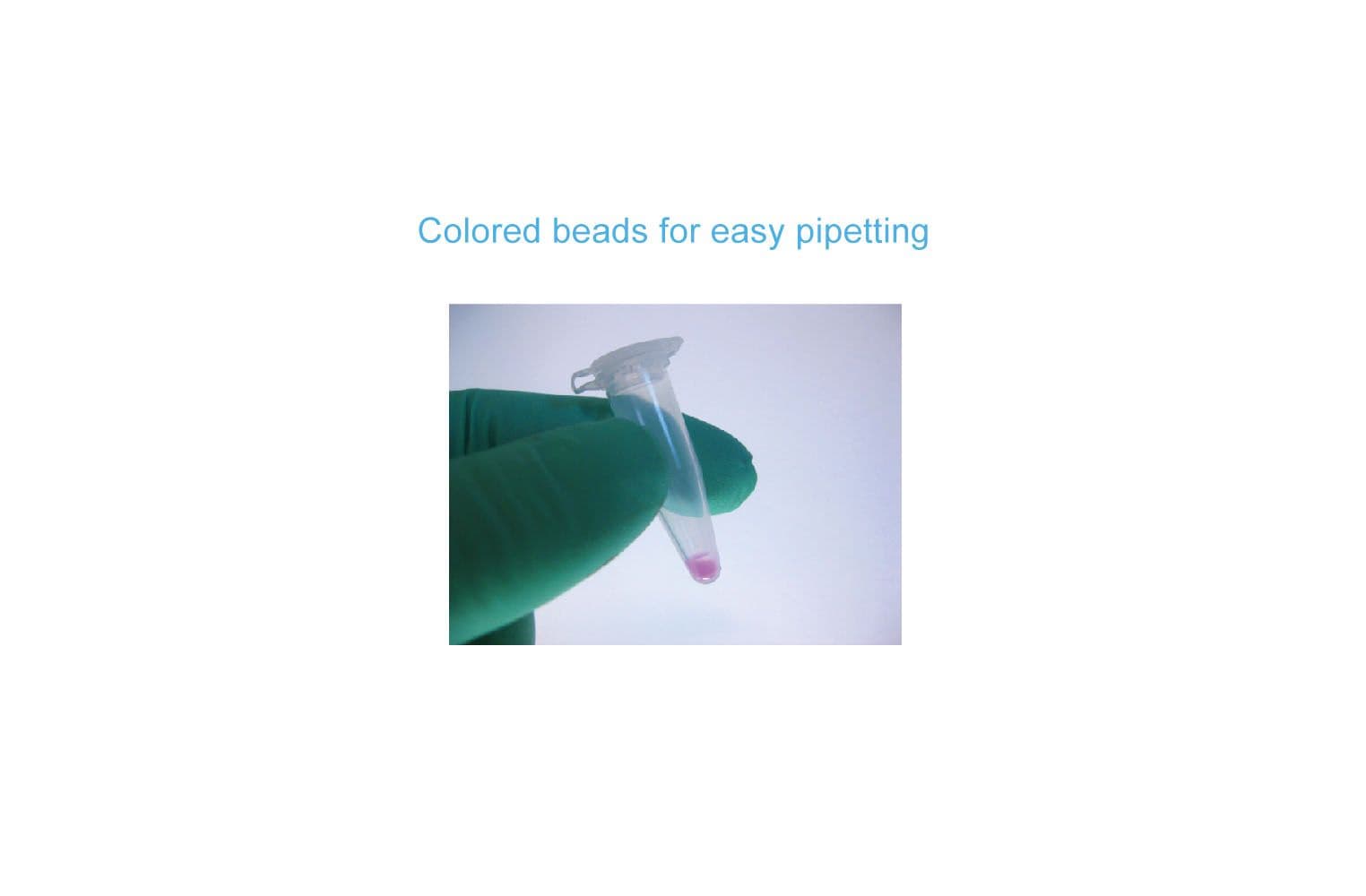
Figure 1.  The brightly colored glutathione agarose beads in BK035 makes the kit easy to use.

Equipment needed

  1. SDS-PAGE minigel system and western blotting transfer apparatus

Example results
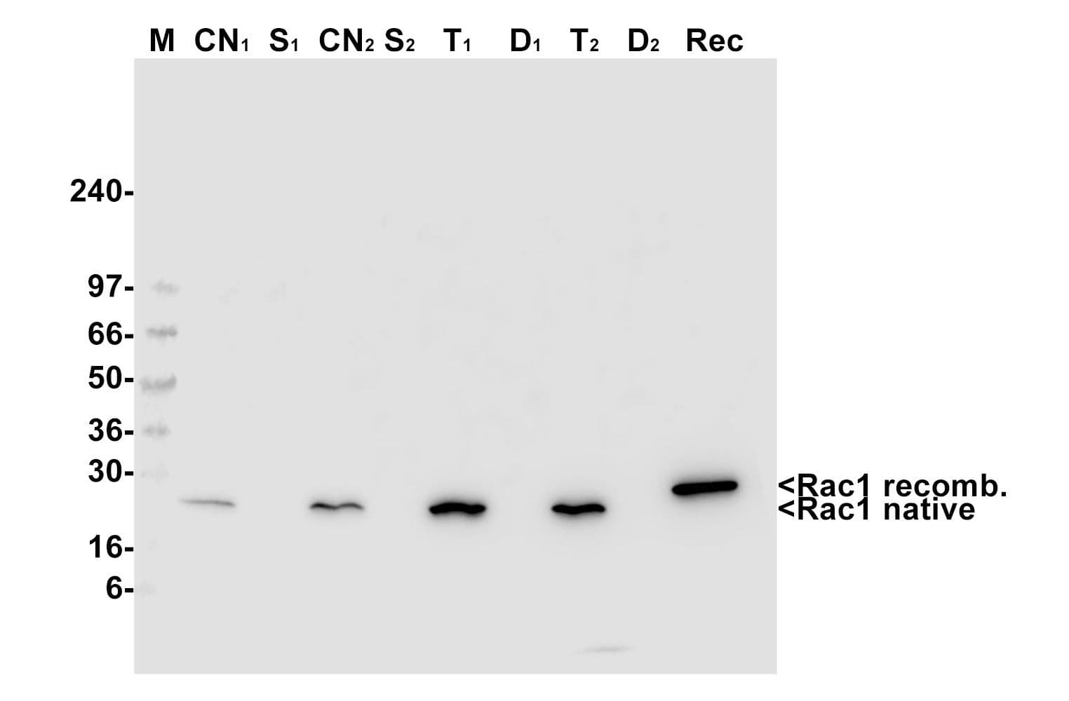
The Rac1 activation assay was tested by loading the Rac1 protein in cell lysates with either GTPγS or GDP. As expected, the GTPγS-loaded Rac1 is very efficiently precipitated while very little GDP-loaded Rac1 is precipitated (Fig. 2).

bk034fig2

Figure 2. Results from BK035 Rac1 activation assay. Activated Rac1 was precipitated and detected in a Western blot using kit BK035. The first lane shows a 50 ng recombinant His-tagged Rac1 standard (Recombinant His-Rac1). The following lanes shows the pull-down of inactive, GDP-loaded Rac1 (Rac1-GDP PD) or active, GTPγS-loaded Rac1 (Rac1-GTP PD) from equal amounts of cell lysates.

Please check out the new version of the Rac Activation Assay and associated products:

G-LISA Products:
Cdc42 G-LISA™ Activation Assay, colorimetric format (Cat.# BK127)
Rac1 G-LISA™ Activation Assay, luminescence format (Cat.# BK126)
Rac1,2,3 G-LISA™ Activation Assay, colorimetric format (Cat.# BK125)
RhoA G-LISA™ Activation Assay, colorimetric format (Cat.# BK124)
RhoA G-LISA™ Activation Assay, luminescence format (Cat.# BK121)

Associated Products:
Anti-Cdc42 monoclonal antibody (Cat.# ACD03)
Anti-Rac1 monoclonal antibody (Cat.# ARC03)
Anti-RhoA monoclonal antibody (Cat.# ARH03)

For product Datasheets and MSDSs please click on the PDF links below.   For additional information, click on the FAQs tab above or contact our Technical Support department at tservice@cytoskeleton.com

AuthorTitleJournalYearArticle Link
Bai, Xiaoyuan et al.The extracellular cyclophilin A-integrin β2 complex as a therapeutic target of viral pneumoniaMolecular Therapy2024
Ramos, Raquel et al.New Insights into the Exosome-Induced Migration of Uveal Melanoma Cells and the Pre-Metastatic Niche Formation in the LiverCancers2024
Ha, Cuong P. et al.Humanin activates integrin αV–TGFβ axis and leads to glioblastoma progressionCell Death & Disease2024
Yau, Eric et al.SP-R210 isoforms of Myosin18A modulate endosomal sorting and recognition of influenza A virus infection in macrophagesMicrobes and Infection2024
Voglewede, Mikayla M. et al.Loss of the polarity protein Par3 promotes dendritic spine neoteny and enhances learning and memoryiScience2024
He, Ling et al.Activation of the Mevalonate Pathway in Response to Anti-cancer Treatments Drives Glioblastoma Recurrences Through Activation of Rac-1Cancer research communications2024
Falace, Antonio et al.FLNA regulates neuronal maturation by modulating RAC1-Cofilin activity in the developing cortexNeurobiology of Disease2024
Laurent-Issartel, Carine et al.Ascites microenvironment conditions the peritoneal pre-metastatic niche to promote the implantation of ovarian tumor spheroids: Involvement of fibrinogen/fibrin and αV and α5β1 integrinsExperimental Cell Research2024
Chen, Ye et al.Dynamic nanomechanical characterization of cells in exosome therapyMicrosystems & Nanoengineering 2024
Guo, Xiong et al.Rabenosyn-5 suppresses non-small cell lung cancer metastasis via inhibiting CDC42 activityCancer Gene Therapy 2024
Yang, Ruicheng et al.Egr-1 is a key regulator of the blood-brain barrier damage induced by meningitic Escherichia coliCell Communication and Signaling2024
Zhang, Huimin et al.PREX1 improves homeostatic proliferation to maintain a naive CD4+ T cell compartment in older ageJCI Insight2024
Veloso, Alexandra et al.The cytoskeleton adaptor protein Sorbs1 controls the development of lymphatic and venous vessels in zebrafishBMC Biology2024
Bahcheli, Alexander T. et al.Pan-cancer ion transport signature reveals functional regulators of glioblastoma aggressionThe EMBO journal2024
Chen, Weiwei et al.Tumour-associated macrophage-derived DOCK7-enriched extracellular vesicles drive tumour metastasis in colorectal cancer via the RAC1/ABCA1 axisClinical and Translational Medicine2024
Aljagthmi, Amjad A. et al.∆Np63α inhibits Rac1 activation and cancer cell invasion through suppression of PREX1Cell Death Discovery2024
Aw, Wen Yih et al.Microphysiological model of PIK3CA-driven vascular malformations reveals a role of dysregulated Rac1 and mTORC1/2 in lesion formationScience Advances2023
Dong, Yuqing et al.CCL2 promotes lymphatic metastasis via activating RhoA and Rac1 pathway and predict prognosis to some extent in tongue cancerCancer Biology & Therapy2023
Takaya, Kento et al.Compound 13 Promotes Epidermal Healing in Mouse Fetuses via Activation of AMPKBiomedicines2023
Koo, Tin Yan et al.N-Acryloylindole-alkyne (NAIA) enables imaging and profiling new ligandable cysteines and oxidized thiols by chemoproteomicsNature Communications2023
Newman, Daniel et al.3D matrix adhesion feedback controls nuclear force coupling to drive invasive cell migrationCell reports2023
Chen, Di et al.Knockdown of Porf-2 restores visual function after optic nerve crush injuryCell Death & Disease2023
Yu, Shuxiang et al.ELMO1 Deficiency Reduces Neutrophil Chemotaxis in Murine PeritonitisInternational Journal of Molecular Sciences2023
Yang, Yuchen et al.β-hydroxybutyrate impairs the directionality of migrating neutrophils through inhibiting the autophagy-dependent degradation of Cdc42 and Rac1 in ketotic cowsJournal of Dairy Science2023
Serwe, Guillaume et al.CNK2 promotes cancer cell motility by mediating ARF6 activation downstream of AXL signallingNature Communications2023
Yau, Eric et al.SP-R210 isoforms of Myosin18A modulate endosomal sorting and recognition of influenza A virus infection in macrophagesMicrobes and Infection2023
Wang, Yueyang et al.Atypical peripheral actin band formation via overactivation of RhoA and nonmuscle myosin II in mitofusin 2-deficient cellseLife2023
Morretton, Jean-Philippe et al.A catalog of numerical centrosome defects in epithelial ovarian cancersEMBO Molecular Medicine2022
Karthikeyan, Subbulakshmi et al.RAB4A GTPase regulates epithelial-to-mesenchymal transition by modulating RAC1 activationBreast Cancer Research2022
Singh, Neeraj et al.BACE-1 inhibition facilitates the transition from homeostatic microglia to DAM-1Science Advances2022
Wang, Yuting et al.EPS8L1 promotes migration and metastasis of ovarian cancer by activating Rac1/MAPK signaling pathway via upregulating TIAM2Authorea Preprints2022
Adan, Hanad et al.Activated Src requires Cadherin-11, Rac, and gp130 for Stat3 activation and survival of mouse Balb/c3T3 fibroblastsCancer Gene Therapy 2022 29:102022
Usuki, Seigo et al.Konjac Ceramide (kCer)-Mediated Signal Transduction of the Sema3A Pathway Promotes HaCaT Keratinocyte DifferentiationBiology2022
Adan, H et al.Activated Src requires Cadherin-11, Rac, and gp130 for Stat3 activation and survival of mouse Balb/c3T3 fibroblastsCancer Gene …2022
Agbaegbu Iweka, Chinyere et al.The lipid phosphatase-like protein PLPPR1 associates with RhoGDI1 to modulate RhoA activation in response to axon growth inhibitory moleculesJournal of Neurochemistry2021
Masi, Ilenia et al.Endothelin-1 drives invadopodia and interaction with mesothelial cells through ILKCell Reports2021
Bai, Xiaoyuan et al.Induction of cyclophilin A by influenza A virus infection facilitates group A Streptococcus coinfectionCell Reports2021
Wang, Jiang Lin et al.Spinophilin modulates pain through suppressing dendritic spine morphogenesis via negative control of Rac1-ERK signaling in rat spinal dorsal hornNeurobiology of Disease2021
Guo, Shuyu et al.Trio cooperates with Myh9 to regulate neural crest-derived craniofacial developmentTheranostics2021
Wang, Junyi et al.Rho-GEF Trio regulates osteosarcoma progression and osteogenic differentiation through Rac1 and RhoACell Death and Disease2021
Zhou, Wenqing et al.Mitofusin 2 regulates neutrophil adhesive migration and the actin cytoskeletonJournal of Cell Science2021
Li, Chunsen et al.GEFT Inhibits Autophagy and Apoptosis in Rhabdomyosarcoma via Activation of the Rac1/Cdc42-mTOR Signaling PathwayFrontiers in Oncology2021
Shin, Seung Kak et al.Exogenous 8-hydroxydeoxyguanosine ameliorates liver fibrosis through the inhibition of Rac1-NADPH oxidase signalingJournal of gastroenterology and hepatology2020
Benz, Peter M. et al.AKAP12 deficiency impairs VEGF-induced endothelial cell migration and sproutingActa physiologica (Oxford, England)2020
Thamilselvan, Vijayalakshmi et al.P-Rex1 Mediates Glucose-Stimulated Rac1 Activation and Insulin Secretion in Pancreatic β-CellsCellular physiology and biochemistry : international journal of experimental cellular physiology, biochemistry, and pharmacology2020
Li, Zean et al.The metastatic promoter DEPDC1B induces epithelial‐mesenchymal transition and promotes prostate cancer cell proliferation via Rac1‐PAK1 signalingClinical and Translational Medicine2020
Moody, Jasmine C. et al.The Rho-GEF PIX-1 directs assembly or stability of lateral attachment structures between muscle cellsNature Communications2020
Danelon, Victor et al.Modular and Distinct Plexin-A4/FARP2/Rac1 Signaling Controls Dendrite MorphogenesisJournal of Neuroscience2020
Larribère, Lionel et al.NF1-RAC1 axis regulates migration of the melanocytic lineageTranslational Oncology2020
Ichikawa, Takehiko et al.Non-junctional role of Cadherin3 in cell migration and contact inhibition of locomotion via domain-dependent, opposing regulation of Rac1Scientific Reports2020
Wei, Yiju et al. NEDD 4L‐mediated Merlin ubiquitination facilitates Hippo pathway activation EMBO reports2020
Aladowicz, Ewa et al.Shcd binds dock4, promotes ameboid motility and metastasis dissemination, predicting poor prognosis in melanomaCancers2020
Wang, Ruixiao et al.Rac1 silencing, NSC23766 and EHT1864 reduce growth and actin organization of bladder smooth muscle cellsLife Sciences2020
Smalley, Tracess et al.The Atypical Protein Kinase C Small Molecule Inhibitor ζ-Stat, and Its Effects on Invasion Through Decreases in PKC-ζ Protein ExpressionFrontiers in Oncology2020
Barabutis, Nektarios et al.Protective mechanism of the selective vas******** V1A receptor agonist selepressin against endothelial barrier dysfunctionJournal of Pharmacology and Experimental Therapeutics2020
Joshi, Rakesh et al.DLC1 SAM domain-binding peptides inhibit cancer cell growth and migration by inactivating RhoAJournal of Biological Chemistry2020
Chen, Lixia et al.CSRP2 suppresses colorectal cancer progression via p130Cas/Rac1 axis-meditated ERK, PAK, and HIPPO signaling pathwaysTheranostics2020
Amato, Clelia et al.WASP Restricts Active Rac to Maintain Cells’ Front-Rear PolarizationCurrent Biology2019
Tsygankova, Oxana M. et al.A unique role for clathrin light chain A in cell spreading and migrationJournal of Cell Science2019
Park, Jin Seok et al.Switch-like enhancement of epithelial-mesenchymal transition by YAP through feedback regulation of WT1 and Rho-family GTPasesNature Communications2019
Lang, Yue et al.MiR-30 family prevents uPAR-ITGB3 signaling activation through calcineurin-NFATC pathway to protect podocytesCell Death and Disease2019
Liu, Yunlong et al.Social Isolation Induces Rac1-Dependent Forgetting of Social MemoryCell Reports2018
McQueeney, Kelley E. et al.Targeting ovarian cancer and endothelium with an allosteric PTP4A3 phosphatase inhibitorOncotarget2018
Zago, Giulia et al.Ralb directly triggers invasion downstream ras by mobilizing the wave complexeLife2018
Barabutis, Nektarios et al.Wild-type p53 enhances endothelial barrier function by mediating RAC1 signalling and RhoA inhibitionJournal of Cellular and Molecular Medicine2018
Shi, Hao et al.Hippo Kinases Mst1 and Mst2 Sense and Amplify IL-2R-STAT5 Signaling in Regulatory T Cells to Establish Stable Regulatory ActivityImmunity2018
Hayashi, Kentaro et al.Intracellular calcium signal at the leading edge regulates mesodermal sheet migration during Xenopus gastrulationScientific Reports2018
Shang, Wanjing et al.Genome-wide CRISPR screen identifies FAM49B as a key regulator of actin dynamics and T cell activationProceedings of the National Academy of Sciences of the United States of America2018
Wang, Yan et al.Involvement of Rac1 signalling pathway in the development and maintenance of acute inflammatory pain induced by bee venom injectionBritish Journal of Pharmacology2016
Han, Jie et al.Farnesyl pyrophosphate synthase inhibitor, ibandronate, improves endothelial function in spontaneously hypertensive ratsMolecular Medicine Reports2016
Wang, Y. et al.Inhibition of prostate smooth muscle contraction and prostate stromal cell growth by the inhibitors of Rac, NSC23766 and EHT1864British Journal of Pharmacology2015
Wang, Shi Jie et al.CD147 promotes Src-dependent activation of Rac1 signaling through STAT3/DOCK8 during the motility of hepatocellular carcinoma cellsOncotarget2015
Hara, Yusuke et al.Directional migration of leading-edge mesoderm generates physical forces: Implication in Xenopus notochord formation during gastrulationDevelopmental Biology2013
Han, Gangwen et al.Preventive and therapeutic effects of Smad7 on radiation-induced oral mucositisNature Medicine2013
Geletu, M. et al.Classical cadherins control survival through the gp130/Stat3 axisBiochimica et Biophysica Acta - Molecular Cell Research2013
Zallocchi, Marisa et al.α1β1 Integrin/rac1-dependent mesangial invasion of glomerular capillaries in alport syndromeAmerican Journal of Pathology2013
Nithipatikom, Kasem et al.Cannabinoid receptor type 1 (CB1) activation inhibits small GTPase RhoA activity and regulates motility of prostate carcinoma cellsEndocrinology2012
Lee, Wonhwa et al.Barrier protective effects of withaferin A in HMGB1-induced inflammatory responses in both cellular and animal modelsToxicology and applied pharmacology2012
Ninkovi, Jana et al.Morphine decreases bacterial phagocytosis by inhibiting actin polymerization through cAMP-, Rac-1-, and p38 MAPK-dependent mechanismsAmerican Journal of Pathology2012
Wong, Hon Kit et al.Merlin/NF2 regulates angiogenesis in schwannomas through a Rac1/semaphorin 3F-dependent mechanismNeoplasia2012
Gastonguay, Adam et al.The role of Rac1 in the regulation of NF-kB activity, cell proliferation, and cell migration in non-small cell lung carcinomaCancer Biology & Therapy2012
Syed, Ismail et al.L-threo-C 6 -pyridinium-ceramide bromide, a novel cationic ceramide, induces NADPH oxidase activation, mitochondrial dysfunction and loss in cell viability in INS 832/13 β-cellsCellular Physiology and Biochemistry2012
Ock, Chan Young et al.A novel approach for stress-induced gastritis based on paradoxical anti-oxidative and anti-inflammatory action of exogenous 8-hydroxydeoxyguanosineBiochemical pharmacology2011
Garkavtsev, Igor et al.Dehydro-α-lapachone, a plant product with antivascular activityProceedings of the National Academy of Sciences of the United States of America2011
Souma, Tomokazu et al.Luminal alkalinization attenuates proteinuria-induced oxidative damage in proximal tubular cellsJournal of the American Society of Nephrology2011
Skalski, Michael et al.SNARE-mediated membrane traffic is required for focal adhesion kinase signaling and Src-regulated focal adhesion turnoverBiochimica et Biophysica Acta - Molecular Cell Research2011
Vidal-Quadras, Maite et al.Rac1 and Calmodulin Interactions Modulate Dynamics of ARF6-Dependent EndocytosisTraffic2011
Balanis, Nikolas et al.β3 integrin-EGF receptor cross-talk activates p190RhoGAP in mouse mammary gland epithelial cellsMolecular Biology of the Cell2011
Jayaram, Bhavaani et al.Isoprenylcysteine carboxyl methyltransferase facilitates glucose-induced Rac1 activation, ROS generation and insulin secretion in INS 832/13 β-cellsIslets2011
McAuley, Erin M. et al.Phenylboronic acid is a more potent inhibitor than boric acid of key signaling networks involved in cancer cell migrationCell Adhesion and Migration2011
Kobayashi, Takashi et al.Activation of Rac1 is closely related to androgen-independent cell proliferation of prostate cancer cells both in vitro and in vivoMolecular Endocrinology2010
Medhora, Meetha et al.20-HETE increases superoxide production and activates NAPDH oxidase in pulmonary artery endothelial cellsAmerican Journal of Physiology - Lung Cellular and Molecular Physiology2008
Bergstrom, R. A. et al.Agrin induced morphological and structural changes in growth cones of cultured hippocampal neuronsNeuroscience2007
Wang, Guoyan et al.Genetic ablation of Rac1 in cartilage results in chondrodysplasiaDevelopmental Biology2007
Slice, Lee W. et al.Angiotensin II and epidermal growth factor induce cyclooxygenase-2 expression in intestinal epithelial cells through small GTPases using distinct signaling pathwaysThe Journal of biological chemistry2005
Sasai, Noriaki et al.The neurotrophin-receptor-related protein NRH1 is essential for convergent extension movementsNature Cell Biol2004
Yang, Seun Ah et al.Rho and Rho-kinase mediate thrombin-induced phosphatidylinositol 4-phosphate 5-kinase trafficking in plateletsJournal of Biological Chemistry2004
Zhang, Yongjun et al.Distinct roles of two structurally closely related focal adhesion proteins, alpha-parvins and beta-parvins, in regulation of cell morphology and survivalThe Journal of biological chemistry2004
Chromy, Brett A. et al.Self-assembly of Abeta(1-42) into globular neurotoxinsBiochemistry2003
Quadri, Sadiqa K. et al.Endothelial Barrier Strengthening by Activation of Focal Adhesion Kinase *Journal of Biological Chemistry2003


Question 1: I have high background and/or multiple bands on my western blot.  How can I fix this?

Answer 1:  There are multiple causes of high background and/or multiple bands.  Some suggestions to improve background signal include:

  1. When blotting use 70v for 45min only as the small G-proteins are very mobile.
  2. Fully remove SDS from the gel by using a non-SDS containing buffer for transfer and performing a full 15 min gel wash step in the transfer buffer before blotting.
  3. Dry the PVDF membrane for 30 min after transfer and before blocking (not necessary for nitrocellulose)
  4. Making sure that the TBST contains 10 mM Tris, 0.05% Tween 20 and 150 mM NaCl.
  5. Incubating with the primary antibody overnight at 4°C and using the appropriate ECL detection system.  


Question 2: How much of the beads should I use for my pull-down experiments?

Answer 2:  PAK-PBD-GST beads (Cat. # PAK02) will bind to Rac1-GDP with a much lower affinity than Rac1-GTP.  If too many PAK-PBD beads are added to the pull-down assay, there will be significant binding to inactive (GDP-bound) Rac1.  The result of this will be an underestimation of Rac1 activation.  For this reason, we highly recommend performing a bead titration to determine optimal conditions for any given Rac1 activation or inactivation assay.  Once optimal conditions have been established, bead titrations should no longer be necessary.  We recommend 10, 15 and 20 μg bead titrations.


Question 3:  How can I test whether the beads are working properly?

Answer 3:  A standard biological assay for PAK-PBD GST protein beads consists of a Rac protein pull-down from cells loaded with either GTPγS (Cat. # BS01) or GDP.  Here are guidelines to follow (see Cat. # PAK02 and BK035 datasheets for more details):


Positive Cellular Protein Control:

Total cell lysate (300 – 800 μg) should be loaded with GTPγS as a positive control for the pull-down assay.  The following reaction details how to load endogenous Rac1 with the nonhydrolysable GTP analog (GTPγS).  This is an excellent substrate for PAK-PBD beads and should result in a strong positive signal in a pull-down assay.


a) Perform GTP loading on 300 – 800 μg of cell lysate (0.5 mg/ml protein concentration) by adding 1/10th volume of Loading Buffer.

b) Immediately add 1/100th volume of GTPγS (200 μM final concentration). Under these conditions 5 - 10% of the Rac1 protein will load with non-hydrolysable GTPγS and will be “pulled-down” with the PAK-PBD beads in the assay.

c) Incubate the control sample at 30°C for 15 min with gentle rotation.

d) Stop the reaction by transferring the tube to 4°C and adding 1/10th volume of STOP Buffer.

e) Use this sample immediately in a pull-down assay.


Negative Cellular Protein Control:

This reaction should be performed in an identical manner to the Positive Control reaction except that 1/100th volume of GDP (1 mM final concentration) should be added to the reaction in place of the GTPγS.  Loading endogenous Rac1 with GDP will inactivate Rac1 and this complex will bind very poorly to PAK-PBD beads.


If you have any questions concerning this product, please contact our Technical Service department at tservice@cytoskeleton.com