Product Uses Include
Studies requiring laminin binding to solid avidin/streptavidin surfaces (1)
Material
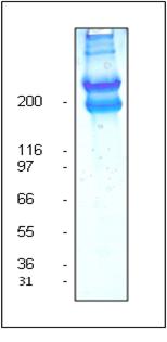
Laminin-1 is purified from EHS tumor tissue and is free of the laminin binding protein entactin which is a common contaminant in some laminin preparations (150 kDa). Protein purity is determined by scanning densitometry of Coomassie Blue stained protein on a 4-20% polyacrylamide gel. The laminin is >90% pure (Figure 1).
The protein is modified to contain covalently linked biotins at random surface lysines. An activated ester of biotin is used to label the protein. The biotin label has been modified to contain a 14 atom spacer between the biotin and the laminin, this improves the accessibility of biotin to avidin/streptavidin.
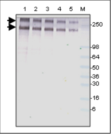
Labeling efficiency is determined by the ability to detect 10 ng of biotinylated laminin using alkaline phosphatase labeled streptavidin (Figure 2). Laminin runs as individual subunits on SDS-PAGE with an apparent molecular weight of 400 and 225 kDa (Figure 1). LMN03 is supplied as an white lyophilized powder. Each vial of LMN03 contains 20 μg protein.
Purity
Purity is determined by scanning densitometry of proteins on SDS-PAGE gels. Samples are >90% pure.
Figure 1: Laminin (biotinylated) purity determination

Legend: Legend: 20 μg of unlabeled laminin was separated by electrophoresis in a 4-20% SDS-PAGE system. The unlabeled protein was stained with Coomassie Blue and visualized in white light. The alpha subunit runs at 400 kDa (top band) while the beta and gamma subunits run as a 225 kDa doublet (lower band). Protein quantitation was determined with the Precision Red™ Protein Assay Reagent (Cat. # ADV02). Mark12 molecular weight markers are from Invitrogen.
Figure 2: Detection of laminin (biotinylated)

Legend: Serial dilutions of biotinylated laminin were separated by electrophoresis on a 4-20% SDS-polyacrylamide gel, blotted to a PVDF membrane, probed with a 1:500 dilution of strepavidin alkaline phosphatase (Sigma) and detected with 1-Step NBY/BCIP reagent™ (Pierce). Lane 1, 100 ng; Lane 2, 50 ng; Lane 3, 40 ng; Lane 4, 20 ng; Lane 5, 10 ng of biotinylated laminin. Lane M, SeeBlue™ molecular weight markers (Invitrogen). Arrows indicate biotinylated laminin subunits (400kD and 225 kD).
|
References
1.Guidebook to the extracellular matrix and adhesion proteins. 1993. Oxford University Press. Ed. Kreis T and Vale R.
For product Datasheets and MSDSs please click on the PDF links below. For additional information, click on the FAQs tab above or contact our Technical Support department at tservice@cytoskeleton.com
If you have any questions concerning this product, please contact our Technical Service department at tservice@cytoskeleton.com
Question 1: What is the size of the spacer between the laminin and the biotin label?
Answer 1: The biotin label has been modified to contain a 14 atom spacer between the biotin and the laminin to improve the accessibility of biotin to avidin/streptavidin.
Question 2: What is the labeling efficiency of the biotinylated laminin?
Answer 2: Labeling efficiency is determined by the ability to detect 10 ng of biotinylated laminin using alkaline phosphatase-labeled streptavidin.
If you have any questions concerning this product, please contact our Technical Service department at tservice@cytoskeleton.com
