Plasma membrane probes
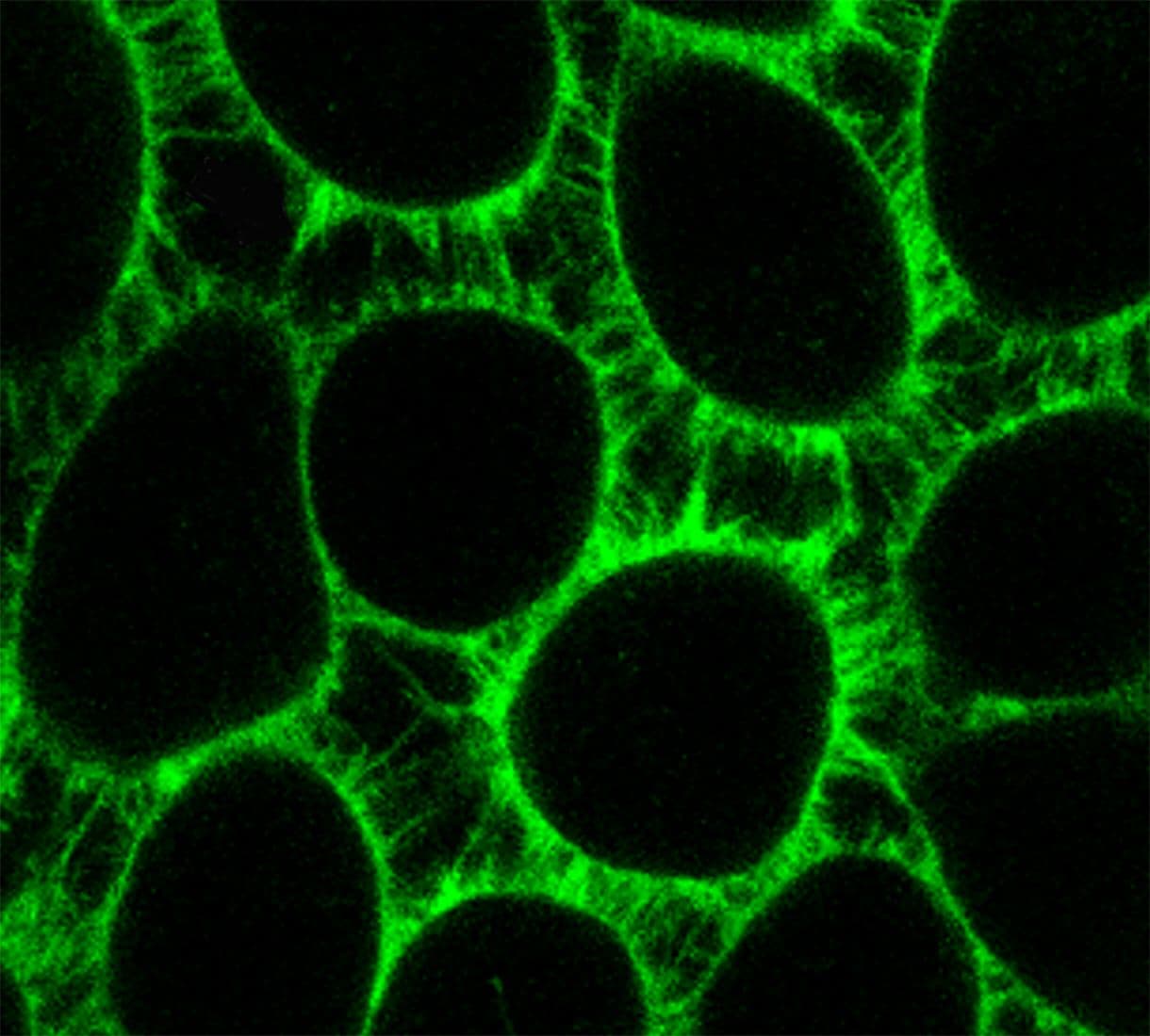
Enhance your membrane visualization with our wide range of MemGlow™ and PK-Mem™ fluorogenic live-cell plasma membrane markers
- Bright and fluorogenic offering exceptionally low background
- Non-phototoxic and non-toxic-allowing long-term imaging and re-imaging of live cells
- Selection of emission spectrum - compatible with multicolor imaging
- Simple staining protocol and low working concentrations
- Does not require any genetic manipulation, transfection or overexpression of fluorescent proteins.
- Compatible with widefield, confocal and many super resolution microscopy formats, including SIM, 2-photon, TIRF, STED.
- Efficient labeling of filopodia and nanotubes at nanomolar concentrations
- No dye quenching or washing steps required
- Most probes are also able to stain fixed cells