The MemGlow™ product line consists of bright & non toxic live cell membrane probes. Originally developed as the MEMBRIGHT™ probes1-3, MemGlow™ fluorogenic probes exhibit ideal microscopy characteristics including high specificity, low background, and simple application. MemGlow™ 560 has been validated with multiple microscopy techniques including epifluorescent (widefield), confocal, 2-photon, and TIRF1. MemGlow™ has been confirmed to work in fixed cells, fixed tissue, live cells, and other phospholipid membranes such as extracellular vesicles including exosomes1-3.
Features and advantages of MemGlow™ probes:
Bright and fluorogenic
Simple staining protocol and low working concentration
Compatible with live and fixed cell staining (see important technical notes in About Tab)
Compatible with ex vivo and fixed tissue staining
Non-toxic probes permit long-term imaging and re-imaging of live cells
Efficient labeling of filopodia and nanotubes at nanomolar concentrations
No dye quenching steps required
Do not alter sample biology
Utilize cyanine or BODIPY dyes with zwitterionic membrane anchor groups
Superior to many existing plasma membrane dyes
For more detailed information on using this product see the About Tab
** 2 nmol of MemGlow will produce a 20µM stock solution when reconstituted with 100 µl of anhydrous DMSO
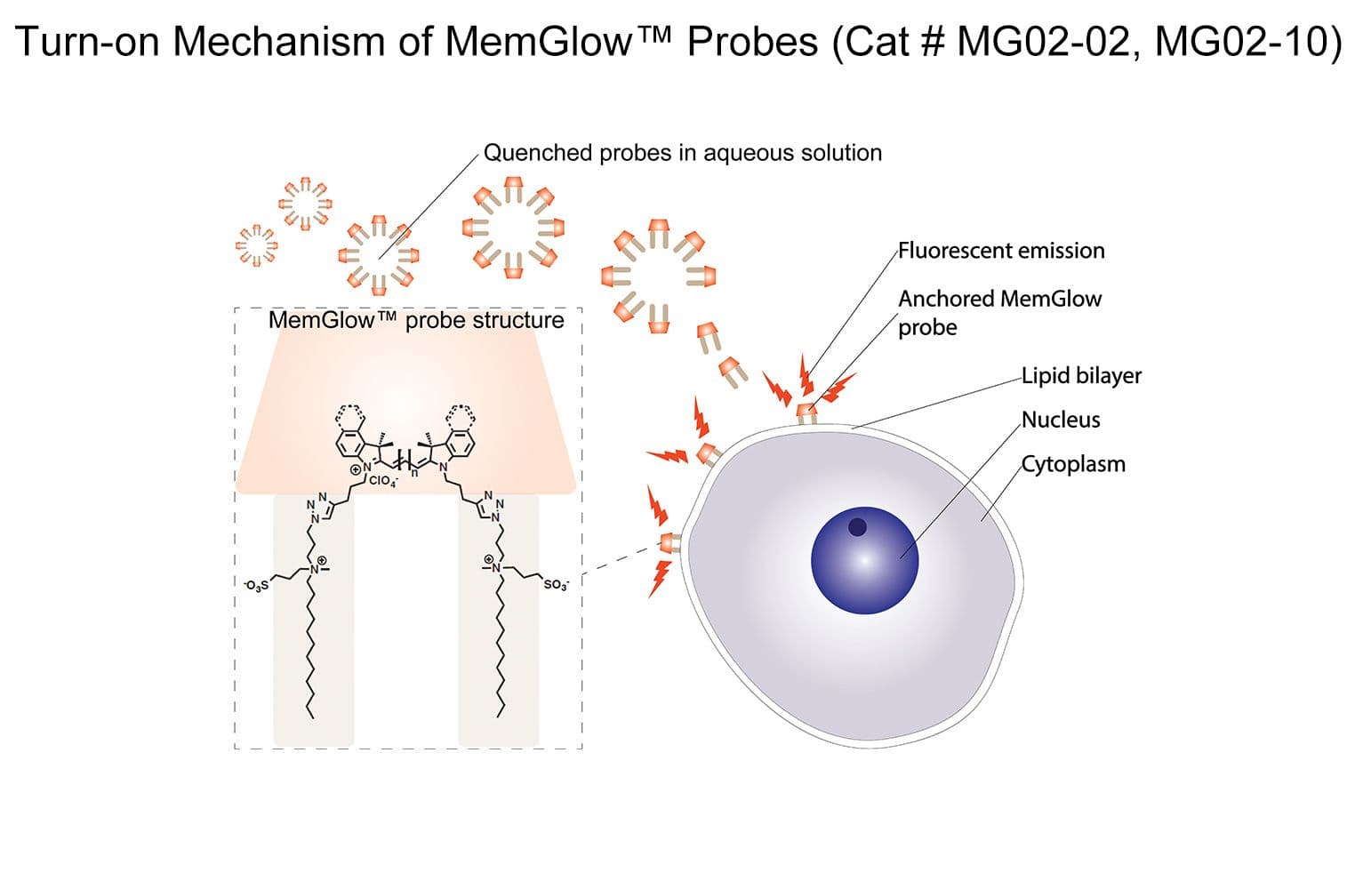
Turn-on mechanism of MemGlow™ probes. MemGlow™ probes are self-quenched nanoparticles until integration with the plasma membrane enables their excitation.
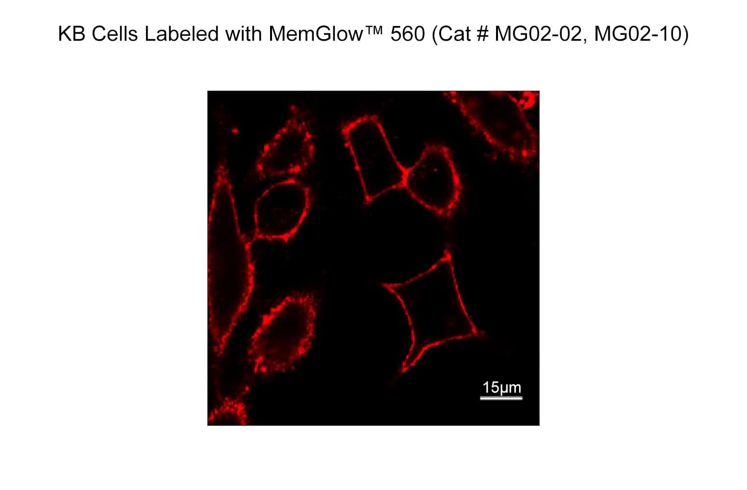
Cytoskeleton's new MemGlow™ probes, part of the MemBright™ family, was used to visualize the plasma membrane of live KB cells with a laser scanning confocal microscope set to 5% laser power (488 nm) for 340 frames with 2 scans per frame and continuous illumination for 15 minutes.
MEMBRIGHT™ is a trademark of CNRS/UNISTRA of France.
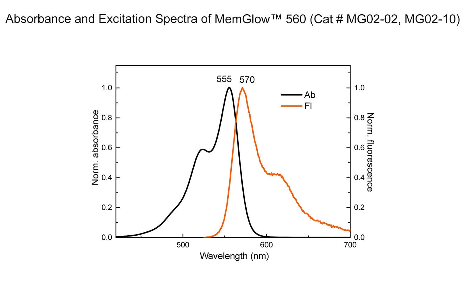
As measured in DMSO, the absorption max of MemGlow™ 560 is 555 nm, with an emission spectra of 570 nm, an extinction coefficient of 150x103, and can be visualized using a tetramethylrhodamine (TRITC) filter set or other suitable filter sets. MemGlow™ 560 is supplied as a lyophilized pellet. Avoid contact with MemGlow™ by wearing appropriate PPE and dispose of according to local regulations and policies.
Storage and Reconstitution
The lyophilized product is stable at 4°C (<10% humidity) for 6 months and should be protected from light. To reconstitute 2 nmol, briefly centrifuge to collect the product at the bottom of the tube. MemGlow™ should be reconstituted with 100 µl of anhydrous DMSO to create a 20 µM stock solution for cell imaging or with 10 µl of anhydrous DMSO to create a 200 µM stock suitable for tissue or small organism imaging. After reconstitution the solution should be stored at -20°C where it is stable for 3 months. Once reconstituted, allow to warm to room temperature before opening tube.
Important Technical Notes
Diluted solutions of MemGlow™ in aqueous media must be used immediately (<20 sec), as MemGlow™ will precipitate and/or bind to tube walls.
Serum can reduce MemGlow™ staining efficiency. When possible MemGlow™ staining should take place in the absence of serum. Optimally, the imaging cell media is serum-free media, reduced serum media, or PBS. In lieu of serum removal, the concentration of MemGlow™ should be increased.
Samples incubating in MemGlow™ solution should be protected from light.
MemGlow™ is non-toxic and live cells can be returned to normal cell media following labeling, and relabeled after 3-4 days.
The localization of MemGlow™ to lipid bilayers is easy to achieve with this product; however, differences in cell morphology and microscope technology, e.g., confocal vs. epifluorescence, will influence the visualization of MemGlow™ (see Figure 3).
When co-labeling with antibodies that require permeabilization limit the concentration of Triton-X to 0.1%.
MemGlow™ is fully compatible with 4% paraformaldehyde (PFA); however, 4% PFA partially permeabilizes the cell membrane so internalization of probes should be expected.
For tissues and small organisms an initial labeling concentration of 2 µM is recommended. For cell culture an initial labeling concentration 20-200 nM is recommended.
Homogeneity of tissue labeling can be optimized with a longer incubation at 4°C rather than relatively brief incubations at room temperature; however, both approaches can label plasma membranes.
Live cells
Fixed cells
Tissue or small organisms
Working Solution (nM)
100
100
2000
Application 1: Labeling the plasma membrane of live cells in culture.
Reagents
MemGlow™ 560 (Cat. # MG02).
Semi-confluent Tib-71 or HEK293 cells grown in a chamber slide.
Imaging medias: PBS, serum-free media or reduced serum media.
Equipment
Fluorescent microscope with a FITC excitation filter at 535 +/-20 nm and emission filter at 585 +/-20 nm for MemGlow™ 560.
Digital camera.
Method
Cells should be seeded onto imaging-appropriate glass or plastics and grown according to cell line requirements to semi-confluency.
Remove any cell culture media from your cells and replace with the media used for imaging (e.g., serum-free media). Do not allow the cells to dry.
Prepare the probe solution by diluting 5 µl of 20 µM MemGlow™ stock in 1 mL imaging media to create a 100 nM working solution or and mix thoroughly. Work quickly (<20 secs) as the probes will begin to aggregate reducing labeling efficiency.
Add diluted probe solution to cells by replacing the cell media with diluted probe solution until covered. Incubate cells in MemGlow™ solution for 10 minutes at room temperature. 37°C incubation can be used but will accelerate endocytosis of probes.
No washing step is required prior to imaging, but can be performed if desired with imaging media.
Proceed with imaging.
Application 2: Labeling the plasma membrane of fixed cells in culture.
Reagents
MemGlow™ 560 (Cat. # MG02).
Semi-confluent Tib71 or HEK293 cells grown on acid-washed coverslips.
Phosphate-buffered saline (PBS, 20 mM potassium phosphate pH 7.4, 150 mM NaCl) .
Fixative solution (4.0 % paraformaldehyde in PBS).
Glass microscope slide.
Coverslip sealing solution (clear nail polish).
EMS Fluoro-Gel mounting media (Cat. # 17985-10)
Equipment
Fluorescent microscope with a FITC excitation filter at 535 +/-20 nm and emission filter at 585 +/-20 nm for MemGlow™ 560.
Digital camera.
Method
Cells should be seeded onto imaging-appropriate glass or plastics and grown according to cell line requirements to semi-confluency.
Remove cell media and wash cells 1X-2X with PBS.
Fix cells for 10-15 minutes at room temperature with 4% paraformaldehyde (PFA).
Remove excess PFA by washing cells with PBS 3X.
(Optional) If co-labeling, permeabilization can be performed at this point. Add 0.1% Triton-X 100 in PBS followed by the primary and secondary antibody protocol according to supplier.
Prepare the probe solution by diluting 5 µl of 20 µM MemGlow™ stock in 1 mL imaging media to create a 100 nM working solution and mix thoroughly. Work quickly (<20 secs) as the probes will begin to aggregate reducing labeling efficiency.
Incubate cells in MemGlow™ solution for 10 minutes at room temperature.
Remove MemGlow™ solution and wash cells with PBS 1X-2X.
If desired place mounting media onto microscope slide.
Apply cover slip cell-side down onto mounting media or microscope slide.
If desired apply coverslip sealing solution according to manufacturers directions.
Proceed with imaging.
Product Citations
Collot, M. et al. MemBright: A Family of Fluorescent Membrane Probes for Advanced Cellular Imaging and Neuroscience. Cell Chem. Biol. 26, 600-614.e7 (2019).
Hyenne, V. et al. Studying the Fate of Tumor Extracellular Vesicles at High Spatiotemporal Resolution Using the Zebrafish Embryo. Dev. Cell 48, 554-572.e7 (2019).
Collot, M., Boutant, E., Lehmann, M. & Klymchenko, A. S. BODIPY with Tuned Amphiphilicity as a Fluorogenic Plasma Membrane Probe. Bioconjug. Chem. 30, 192–199 (2019).
MEMBRIGHT™ is a trademark of CNRS/UNISTRA of France.
Q1. Can I use MemGlow™ in experiments that require cell permeabilization for antibody co-labeling?
A1. Yes, MemGlow™ have been successfully used in co-labeling experiments requiring permeabilization using Triton-X 100 up to 0.1%¹.
Q2. Is MemGlow™ compatible with live cell labeling?
A2. Yes, MemGlow™ is non-toxic and can be used to label live cells¹. After imaging, labeled cells can be returned to normal cell media and relabeled again 3-4 days later.
Q3. Will MemGlow™ efficiently label tissues or other lipid bilayers such exosomes?
A3. Yes, MemGlow™ has been used to label fixed and sectioned tissues¹, ex vivo liver tissues¹, and exosomes³.
Q4. Will MemGlow™ produce precise localization of the plasma membrane in fixed cell applications?
A4. MemGlow™ localizes specifically to the plasma membrane of cells; however, if the cell membrane is disrupted, MemGlow will label phospholipids within the cellular milieu. As paraformaldehyde fixation partially permeabilizes the plasma membrane, intracellular MemGlow signal should be expected.
Raw 267.4 cell fixed with 4% PFA and labeled with MemGlow. MemGlow signal is seen throughout the cell.
Citations
Collot, M. et al. MemBright: A Family of Fluorescent Membrane Probes for Advanced Cellular Imaging and Neuroscience. Cell Chem. Biol. 26, 600-614.e7 (2019).
Collot, M., Boutant, E., Lehmann, M. & Klymchenko, A. S. BODIPY with Tuned Amphiphilicity as a Fluorogenic Plasma Membrane Probe. Bioconjug. Chem. 30, 192–199 (2019).
Hyenne, V. et al. Studying the Fate of Tumor Extracellular Vesicles at High Spatiotemporal Resolution Using the Zebrafish Embryo. Dev. Cell 48, 554-572.e7 (2019).