Flipper-TR™ membrane tension probes
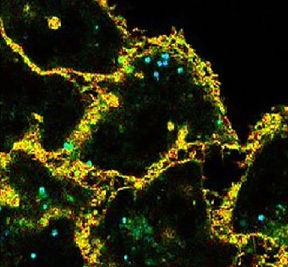
Changes in membrane tension play a central role in numerous cellular processes, including motility, cell division, phagocytosis, and endocytosis. The introduction of the Flipper-TR™ line of fluorogenic probes has dramatically simplified what was once a highly technical and labor-intensive process. These innovative probes, designed for live-cell imaging using the Fluorescent Lifetime Imaging Microscopy (FLIM) technique, provide a convenient way to visualize dynamic changes in membrane tension. The Flipper-TR™ probes are available for targeting the plasma membrane, mitochondria, and lysosomes, enabling you to study membrane mechanics across multiple organelles with ease.
Also available is the versatile Halo-Flipper reagent, which precisely localizes the Flipper-TR probe to your Halo-tagged protein of interest (POI) within cells and senses changes in the organization of lipid bilayer membranes surrounding your POI.