About HAK-actin™
HAK-actin™ is a pan-species Expansion Microscopy (ExM) compatible probe for actin visualization using U-ExM based protocols. HAK-actin™ binds specifically to F-actin. Its design allows HAK-actin™ to be anchored into the gel and expand faithfully to the original actin structure after the swelling step. HAK-actin™ contains an HA tag epitope, it can therefore be detected post expansion using a pair of anti-HA and species specific secondary antibodies. It combines simplicity through a single protocol and versatility as the fluorophore label on the secondary antibody can be chosen freely. Finally, the primary + secondary antibody boosts fluorescent signal amplification, circumventing volumetric dilution of the fluorescent signal due to expansion.
HAK-actin
Cat. # CY-SC019

HeLa cells stained with HAK-actin, on the right showing depth as a color spectrum.
Samples were fixed and processed for expansion microscopy as detailed in Mercey O. et al. 2025. After two 5 min washes in PBS, samples were expanded (using 3 rounds of H2O), and incubated with rabbit-anti HA (1/340 dilution, orange on left) in blocking buffer plus SPY650-DNA for 3.5 h at RT. After 3 washes in PBS PBS/0.2% TritonX100, they were incubated with secondary antibody (1/500 dilution) in blocking buffer for 90 min at RT in the dark.
Gels were then washed in PBS and PBS/0.2% TritonX100 and mounted in anti-fade media.
Mercey O. et al 2025. HAK-actin, U-ExM-compatible probe to image the actin cytoskeleton. https://www.biorxiv.org/content/10.1101/2025.08.26.672318v1.
How does HAK-actin™ work?
The HAK-actin™ probe contains 3 key elements:
1. A pan-species, highly specific, high affinity F-actin ligand for a robust binding to polymerized actin (F-actin) only.
2. An anchoring moiety that specifically reacts with acrylamide and covalently links HAK-actin™ to the expansion gel.
3. The HA-tag epitope sequence for a strong and specific recognition by anti-HA antibodies, even after expansion.

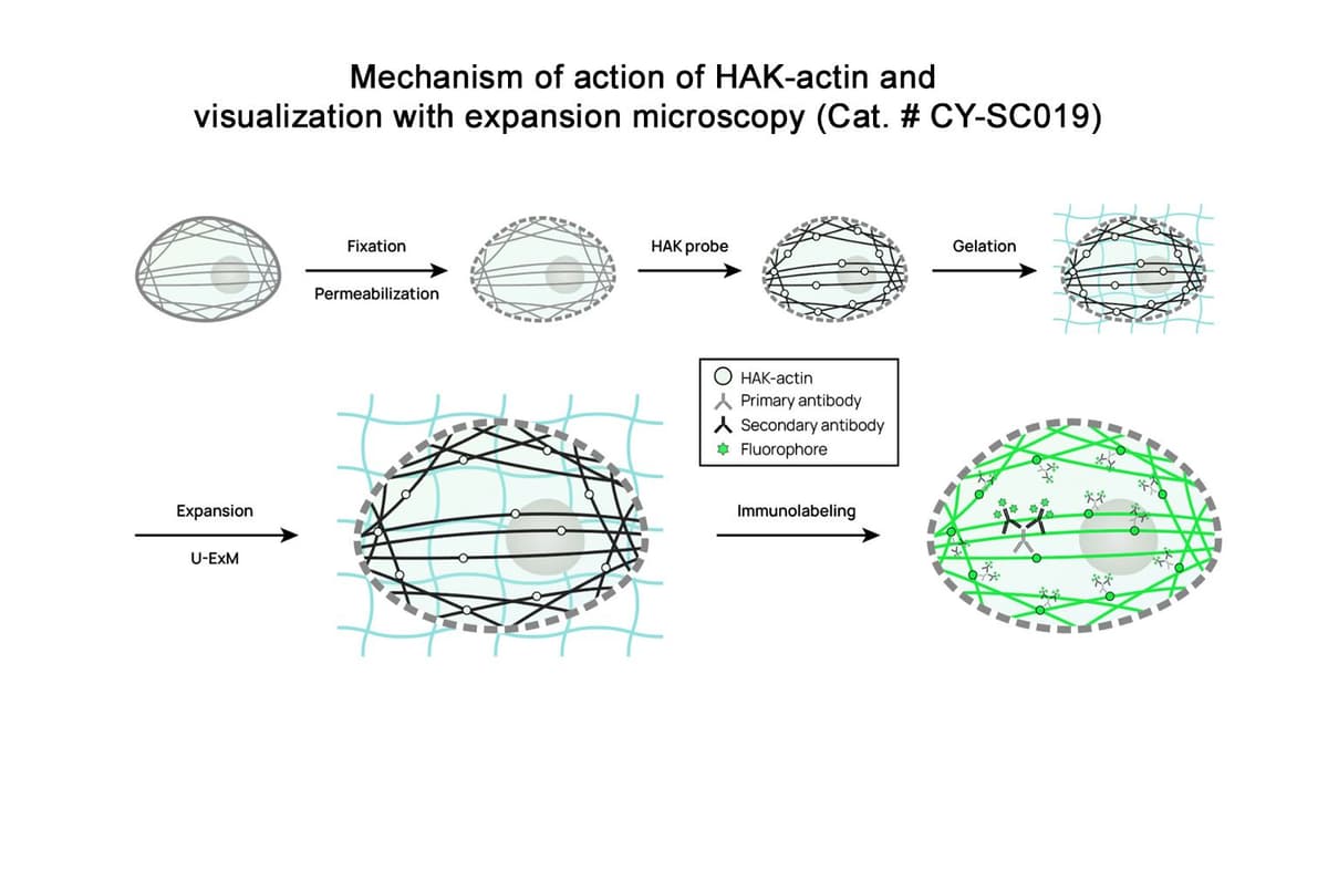
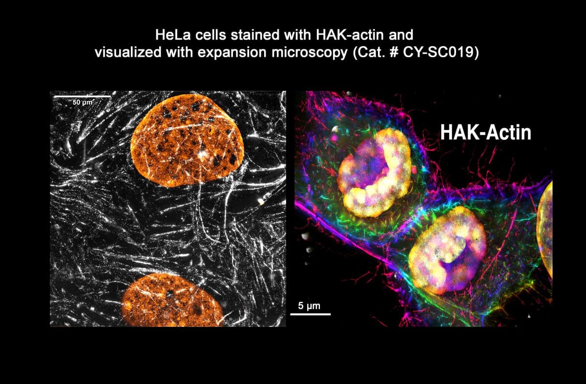
Figure 1. After fixation (chemical or cryo) and permeabilization, the sample to be expanded is treated with HAK-actin™. The probe’s actin ligand uniformly labels F-actin within the cells. The sample is then subjected to U-ExM (or iU-ExM) protocol. During the protocol, HAK-actin™ is covalently bound within the expansion gel and expands faithfully with the sample. The final immunolabeling step using a primary anti-HA and a fluorescently labeled secondary antibody boosts the fluorescent signal and reveals the expanded actin structure with high contrast.
Stronger signal via post-expansion immunostaining. The HA tag allows post-U-ExM labeling with standard anti-HA antibodies, giving ~4× higher fluorescence than a directly labeled probe. The user is free to choose any fluorophore on the secondary antibody.
Robust anchoring/retention in the gel. Four lysines promote efficient crosslinking to the U-ExM polymer, yielding bright, specific actin staining with low background.
Wide protocol compatibility. Works with standard U-ExM, cryo-ExM (better ultrastructure preservation), and iterative U-ExM (up to ~16× expansion), and can be multiplexed with other labels (e.g., Microtubules or membranes with mCLING).
Broad applicability across models. Delivers uniform actin labeling in human cells (U2OS), neurons (growth cones, dendritic spines), platelets, mouse retina (clearer signal in the ciliary bulge), and diverse microbial eukaryotes—including cell-walled species where anti-actin or phalloidin often fail.
Overcomes key ExM limitations. By using post-labeling, it avoids the fluorophore dilution and color constraints of pre-labeling approaches (e.g., AcX-based phalloidin linkers), doesn’t require species-specific antibodies or genetic tagging, and simplifies a reproducible workflow.
No transfection or expression required. HAK-actin does not require genetic expression of tagged actin monomers. It labels endogenous actin.
Note: HAK-actin™ has been specifically designed for U-ExM, iU-ExM and Cryo-ExM protocols, other ExM methods might not work. The detailed protocols of these compatible expansion microscopy methods can be found in the following publications:
U-ExM: https://doi.org/10.1016/bs.mcb.2020.05.006