Acti-stain 670 phalloidin

Acti-stain 670 phalloidin
$0.00

Product Uses Include

    • Fluorescent staining of fixed actin filaments in tissue sections and tissue culture cell preparations.Note: Unlike many actin antibodies, Acti-stain™ 670 phalloidin binds only to F-actin resulting in low background fluorescence.  Furthermore, actin staining is not appreciably different between species.
    • Preparation of stabilized fluorescent actin filaments in vitro.


    Actin staining is very useful in determining the structure and function of the cytoskeleton in living and fixed cells. The actin cytoskeleton is a very dynamic and labile structure in the living cell, but it can be fixed with paraformaldehyde prior to probing or staining for actin structures. 


    Material

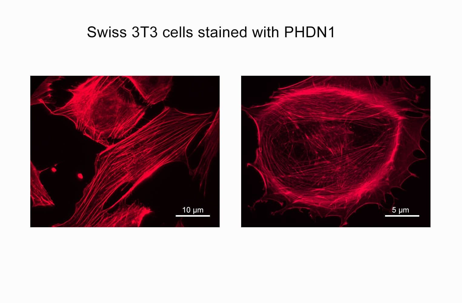
    Phalloidin is a seven amino acid peptide toxin from the mushroom Amanita phalloides, which binds specifically and with high affinity (Kd 20 nM) to the polymerized form of actin (F-actin).  Phalloidin lowers the critical concentration of actin polymerization to less than 1 µg/ml, thereby acting as a polymerization enhancer. Phalloidin has been labeled with a proprietary far-red fluorescent dye which allows it to be used to stain actin filaments in tissue cultured cells and tissue sections (1, see Figure 1) and cell-free preparations.  Acti-stain™ 670 phalloidin-labeled actin filaments retain many functional characteristics of unlabeled actin including their ability to interact with myosin.  Actin-stain™ 670 phalloidin is supplied as a light blue solid. 

    Note: Phalloidin is toxic and must be handled with care (LD50 human = 2mg/Kg).


    Example Results and Specifications

    Figure 1.   Actin Stress Fibers stained with Acti-stain™ 670 in a Swiss 3T3 cell.

    PHDN1_img1

    Mitotic Swiss 3T3 cell, F-actin stained with Acti-stain™ 670 (far-red, Cat. # PHDN1), nuclear DNA stained with Dapi (blue).


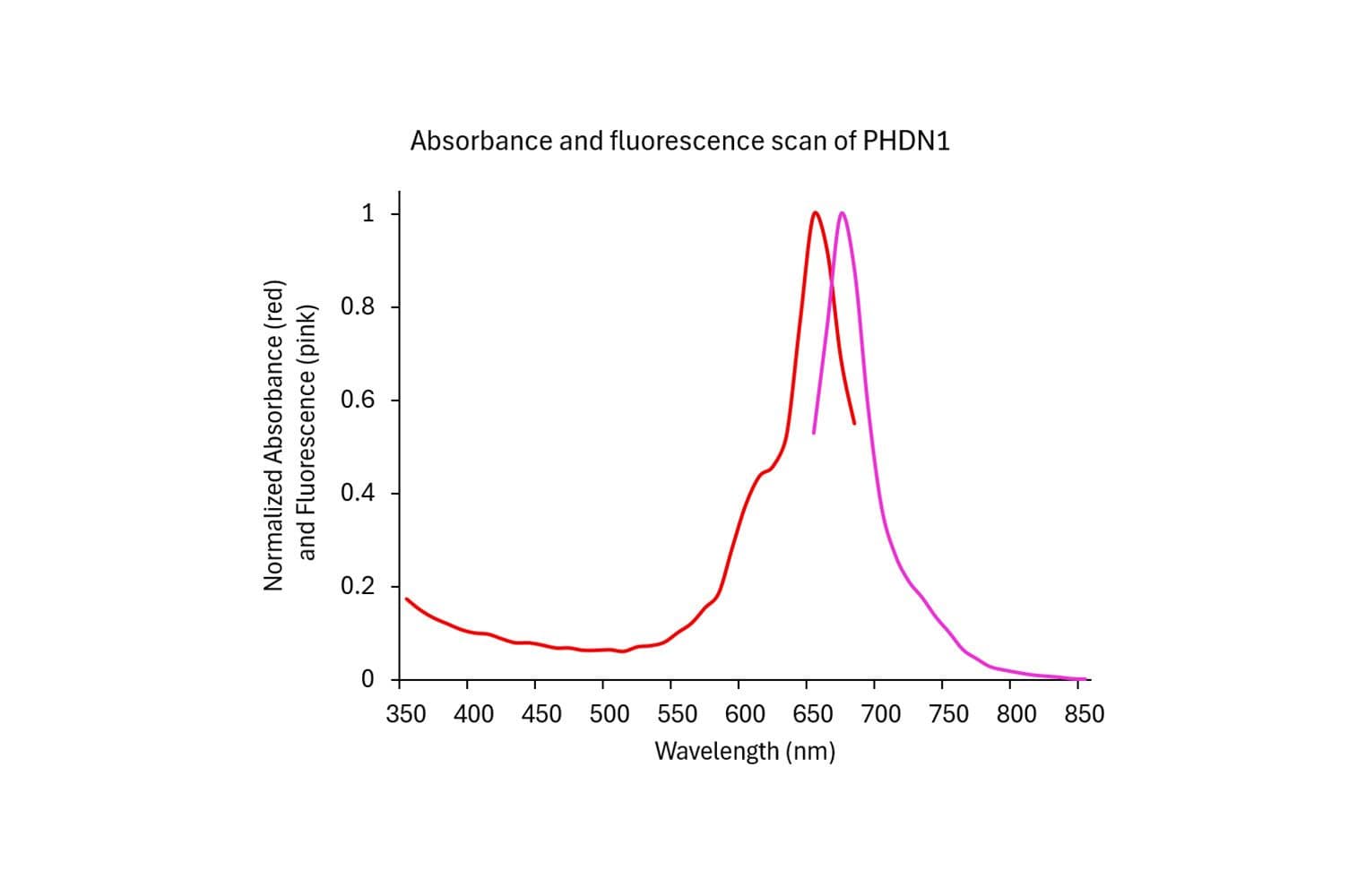
    Figure 2. Emission and excitation scans for Acti-stain™ phalloidins

    PHDN1_scan

    Absorbance and fluorescence scan of Acti-stain™ 670. Labeled phalloidin was diluted into methanol and its absorbance and excitation spectra were scanned between 300-750 and 600-750 nm, respectively. Absorbance peaks at 625 nm and fluorescence at 675 nm.

    Acti-stain phalloidins are the most well characterized phalloidins available. Tabe 1 describes their brightness, photostability, background and affinity constants to F-actin. Compare these performance characteristics to other fluorescent phalloidins and you will see the advantages of using Acti-stain™ for your actin staining requirements.

    Table 1. Biochemical characteristics of fluorescent phalloidins

    ConjugateCat.#

    Wavelengths (Ex/Em)

    Brightness (AFU*)

    Stability to photobleaching(half life in seconds**)

    Background (% of total AFU at 100nM**)

    Affinity (Kd in nanomolar)

    Fluorescein-phalloidin

    na485/535 FITC filter set43262272 +/-12

    Acti-stain™ 488

    PHDG1485/535 FITC filter set83257555 +/-8

    Acti-stain™ 535

    PHDR1535/585 TRITC filter set430271236 +/-7

    Acti-stain™ 555

    PHDH1535/585 TRITC filter set551461663 +/-8

    Acti-stain™ 670

    PHDN1640/680 Cy5 filter set332181850 +/-12

    * = AFU's measured by quantitative cell imaging. ** = Measured in stained Swiss 3T3 cells in the absence of antifade.


    For more information about protocols to fix and stain cells click here.


    References

    1. Wulf, E. et al. (1979). Proc Natl Acad Sci USA. 76(9):4498-4502.

    2. Kron, S.J. et al. (1991).  Meth. Enzmol. 196: 399-416.

    For product Datasheets and MSDSs please click on the PDF links below.   For additional information, click on the FAQs tab above or contact our Technical Support department at tservice@cytoskeleton.com

    AuthorTitleJournalYearArticle Link
    Quadri, Zainuddin et al.Ceramide-mediated orchestration of oxidative stress response through filopodia-derived small extracellular vesiclesJournal of Extracellular Vesicles2024
    Bekedam, Fjodor T. et al.Mechanical stimulation of induced pluripotent stem cell derived cardiac fibroblastsScientific Reports2024
    Cheng, Ya Wen et al.Shear Stress and Sub-Femtomolar Levels of Ligand Synergize to Activate ALK1 Signaling in Endothelial CellsCells2024
    Bhosle, Vikrant K. et al.The chemorepellent, SLIT2, bolsters innate immunity against Staphylococcus aureuseLife2023
    Karvas, Rowan M. et al.3D-cultured blastoids model human embryogenesis from pre-implantation to early gastrulation stagesCell stem cell2023
    Gromova, Kira V. et al.The kinesin Kif21b binds myosin Va and mediates changes in actin dynamics underlying homeostatic synaptic downscalingCell reports2023
    Fu, Jing et al.The checkpoint inhibitor PD-1H/VISTA controls osteoclast-mediated multiple myeloma bone diseaseNature Communications2023
    Newman, Daniel et al.3D matrix adhesion feedback controls nuclear force coupling to drive invasive cell migrationCell reports2023
    Isomursu, Aleksi et al.Dynamic Micropatterning Reveals Substrate-Dependent Differences in the Geometric Control of Cell Polarization and MigrationSmall Methods2023
    Ferrari, Dario et al.Effects of biomechanical and biochemical stimuli on angio- and vasculogenesis in a complex microvasculature-on-chipiScience2023
    Podieh, Fabienne et al.Differential role for rapid proteostasis in Rho GTPase-mediated control of quiescent endothelial integrityJournal of Biological Chemistry2023
    Sengupta, Arunima et al.A New Immortalized Human Alveolar Epithelial Cell Model to Study Lung Injury and Toxicity on a Breathing Lung-On-Chip SystemFrontiers in Toxicology2022
    Cobham, Ansa E. et al.Maintaining robust size across environmental conditions through plastic brain growth dynamicsOpen Biology2022
    Gau, David et al.Myocardin‐related transcription factor's interaction with serum‐response factor is critical for outgrowth initiation, progression, and metastatic colonization of breast cancer cellsFASEB BioAdvances2022
    Karvas, Rowan M. et al.Stem-cell-derived trophoblast organoids model human placental development and susceptibility to emerging pathogensCell Stem Cell2022
    Basu, Arkaprabha et al.Statistical parametrization of cell cytoskeleton reveals lung cancer cytoskeletal phenotype with partial EMT signatureCommunications Biology 2022
    Chen, Jia Wei et al.mito-TEMPO Attenuates Oxidative Stress and Mitochondrial Dysfunction in Noise-Induced Hearing Loss via Maintaining TFAM-mtDNA Interaction and Mitochondrial BiogenesisFrontiers in Cellular Neuroscience2022
    Woo, Marcel S. et al.Neuronal metabotropic glutamate receptor 8 protects against neurodegeneration in CNS inflammationJournal of Experimental Medicine2021
    Schimmel, Lilian et al.Endothelial cells are not productively infected by SARS-CoV-2Clinical and Translational Immunology2021
    Goering, Jeremy P. et al.In-frame deletion of SPECC1L microtubule association domain results in gain-of-function phenotypes affecting embryonic tissue movement and fusion eventsHuman molecular genetics2021
    Majolée, Jisca et al.The interplay of Rac1 activity, ubiquitination and GDI binding and its consequences for endothelial cell spreadingPLoS ONE2021
    Thillaiappan, Nagendra Babu et al.KRAP tethers IP3 receptors to actin and licenses them to evoke cytosolic Ca2+ signalsNature Communications2021
    Merckaert, Tijs et al.An AKT2-specific nanobody that targets the hydrophobic motif induces cell cycle arrest, autophagy and loss of focal adhesions in MDA-MB-231 cellsBiomedicine and Pharmacotherapy2021
    Schiweck, Juliane et al.Drebrin controls scar formation and astrocyte reactivity upon traumatic brain injury by regulating membrane traffickingNature Communications2021
    Chen, Lena et al.Ankyrin-B p.S646F undergoes increased proteasome degradation and reduces cell viability in the H9c2 rat ventricular cardiomyoblast cell lineBiochem Cell Biol2020
    Chemla, Yoav et al.Carbon nanostructures as a scaffold for human embryonic stem cell differentiation toward photoreceptor precursorsNanoscale2020
    Peron-Cane, Caroline et al.Fluorescent secreted bacterial effectors reveal active intravacuolar proliferation of Listeria monocytogenes in epithelial cellsPLoS Pathogens2020
    Huang, Rui et al.NCAM regulates temporal specification of neural progenitor cells via profilin2 during corticogenesisJournal of Cell Biology2020
    Vohnoutka, Rishel B. et al.The focal adhesion scaffold protein Hic-5 regulates vimentin organization in fibroblastsMolecular Biology of the Cell2019
    Jung, Wei Hung et al.Cell–cell adhesion and Myosin activity regulate cortical actin assembly in mammary gland epithelium on concaved surfaceCells2019
    Han, Yuhong et al.Phosphorylation of Ci/Gli by Fused Family Kinases Promotes Hedgehog SignalingDevelopmental Cell2019
    Majolée, Jisca et al.CSN5 inhibition triggers inflammatory signaling and Rho/ROCK-dependent loss of endothelial integrityScientific Reports2019
    Marques, Pedro E. et al.Multimerization and Retention of the Scavenger Receptor SR-B1 in the Plasma MembraneDevelopmental Cell2019
    Srikanth, Priya et al.Convergence of independent DISC1 mutations on impaired neurite growth via decreased UNC5D expressionTranslational Psychiatry2018
    Bertier, Laurence et al.Nanobodies targeting cortactin proline rich, helical and actin binding regions downregulate invadopodium formation and matrix degradation in SCC-61 cancer cellsBiomedicine and Pharmacotherapy2018
    Bertier, Laurence et al.Inhibitory cortactin nanobodies delineate the role of NTA- and SH3-domain-specific functions during invadopodium formation and Cancer cell invasionFASEB Journal2017
    Rolland-Fourcade, Claire et al.Epithelial expression and function of trypsin-3 in irritable bowel syndromeGut2017
    Parker, Christopher G. et al.Ligand and Target Discovery by Fragment-Based Screening in Human CellsCell2017
    Osborne, Suzanne E. et al.Type I interferon promotes cell-to-cell spread of Listeria monocytogenesCellular Microbiology2017
    Dombert, Benjamin et al.BDNF/trkB Induction of Calcium Transients through Cav2.2 Calcium Channels in Motoneurons Corresponds to F-actin Assembly and Growth Cone Formation on β2-Chain Laminin (221)Frontiers in Molecular Neuroscience2017
    Erdogan, Begum et al.Cancer-associated fibroblasts promote directional cancer cell migration by aligning fibronectinJournal of Cell Biology2017
    Mi, Na et al.CapZ regulates autophagosomal membrane shaping by promoting actin assembly inside the isolation membraneNature Cell Biology2015
    Shimamura, Shintaro et al.The Src substrate SKAP2 regulates actin assembly by interacting with WAVE2 and cortactin proteinsJournal of Biological Chemistry2013
    Cuajungco, Math P. et al.Abnormal accumulation of human transmembrane (TMEM)-176A and 176B proteins is associated with cancer pathologyActa Histochemica2012
    Ahrens, Susan et al.F-Actin Is an Evolutionarily Conserved Damage-Associated Molecular Pattern Recognized by DNGR-1, a Receptor for Dead CellsImmunity2012

    Question 1:  Can I use fluorescently-labeled phalloidin to stain F-actin in living cells?

    Answer 1:  Unfortunately, no, phalloidin cannot be used to stain F-actin in living cells.  Phalloidins are used to stain F-actin in fixed cells.  Fluorescent phalloidins only bind to the native quaternary structure of F-actin which provides a low background.  The correct fixation condition for phalloidin binding is 3.7% (v/v) paraformaldehyde in PBS for 10 min because it retains the quaternary protein structure which is necessary for high affinity binding of phalloidin.  Methanol fixation destroys the native conformation and hence is not suitable for F-actin staining with phalloidin.  To monitor actin dynamics in living cells, micro-injection of rhodamine-labeled actin (Cat. # APHR or AR05) is recommended.  Please see those datasheets for more information.


    Question 2:  Which of the fluorescently-labeled phalloidin is the most stable/brightest?

    Answer 2:  The brightest and most stable of the Acti-stains is Acti-stain 488 (green fluorescence; Cat. # PHDG1).  Please see the table below for additional information on all of our Acti-stains. 

    Conjugate

    Cat. #

    Wavelengths (Ex/EM)

    Brightness (AFU)

    Stability to photobleaching* (1/2 life in sec)

    Background (% of total AFU at 100 nM)

    Acti-stain™ 488

    PHDG1

    485/535

    832

    57

    5

    Acti-stain™ 535

    PHDR1

    535/585

    430

    27

    12

    Acti-stain™ 555

    PHDH1

    535/585

    551

    46

    16

    Acti-stain™ 670

    PHDN1

    640/680

    332

    18

    18

    * = measured in the absence of antifade.


    If you have any questions concerning this product, please contact our Technical Service department at tservice@cytoskeleton.com.