Cdc42 Pull-down Activation Assay Biochem Kit (bead pull-down format) - 50 Assays

Cdc42 Activation Assay Biochem Kit (bead pull down format) - 50 Assays
$0.00

Product Uses Include

  • Analysis of in vivo Cdc42 activation.

Introduction
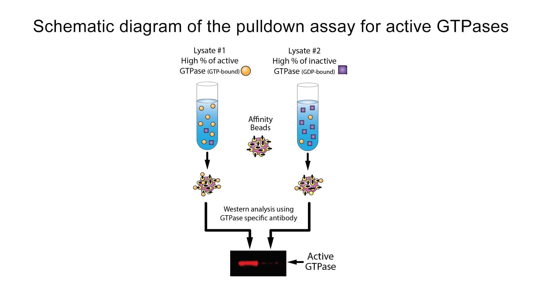
The Rho switch operates by alternating between an active, GTP-bound state and an inactive, GDP-bound state.  Understanding the mechanisms that regulate activation / inactivation of the GTPases is of obvious biological significance and is a subject of intense investigation.  The fact that many Rho family effector proteins will specifically recognize the GTP bound form of the protein has been exploited experimentally to develop a powerful affinity purification assay that monitors Rac and Cdc42 protein activation.  The assay uses the Cdc42/Rac Interactive Binding (CRIB) region (also called the p21 Binding Domain, PBD) of the Cdc42 / Rac effector protein, p21 activated kinase 1 (PAK).  The CRIB/PBD protein motif has been shown to bind specifically to the GTP-bound form of Rac and/or Cdc42 proteins.  The fact that the PBD region of PAK has a high affinity for both GTP-Rac and GTP-Cdc42 and that PAK binding results in a significantly reduced intrinsic and catalytic rate of hydrolysis of both Rac and Cdc42 make it an ideal tool for affinity purification of GTP-Rac and GTP-Cdc42 from cell lysates.  The PAK-PBD protein supplied in this kit corresponds to residues 67-150.  This includes the highly conserved CRIB region (aa 74-88) plus sequences required for the high affinity interaction with GTP-Rac and GTP-Cdc42.  The PAK-PBD is in the form of a GST fusion protein, which allows one to "pull-down" the PAK-PBD/GTP-Cdc42 (or GTP-Rac) complex with glutathione affinity beads.  The assay therefore provides a simple means of quantitating Rac/Cdc42 activation in cells.  The amount of activated Cdc42 is determined by a Western blot using a Cdc42 specific antibody.


Kit contents
The kit contains sufficient materials for 50 assays, depending on assay setup, and includes reagents for positive and negative controls. The following components are included:

  1. GST-tagged PAK-PBD protein on colored agarose beads (Cat. # PAK02)
  2. Cdc42 monoclonal antibody (Cat. # ACD03)
  3. His-tagged Cdc42 protein (Cat. # CD01)
  4. GTPγS: (non-hydrolyzable GTP analog) (Cat. # BS01)
  5. GDP
  6. Cell lysis Buffer
  7. Wash Buffer
  8. Loading Buffer
  9. STOP Buffer
  10. Protease inhibitor cocktail (Cat. # PIC02)
  11. Manual with detailed protocols and extensive troubleshooting guide
beads

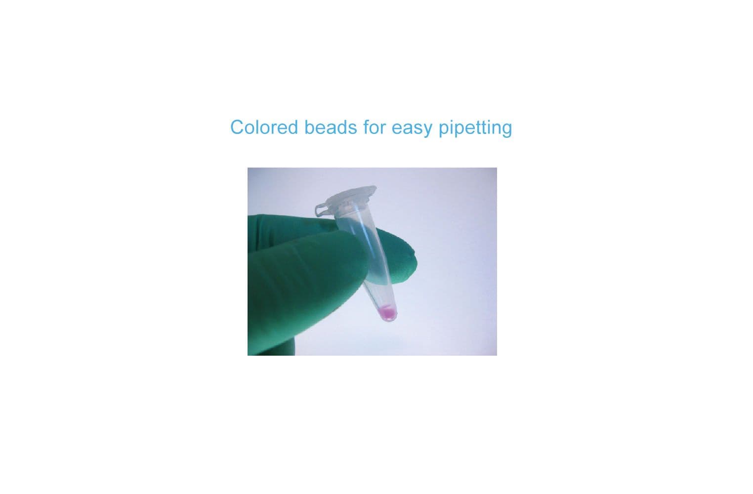
Figure 1. The brightly colored glutathione agarose beads in BK034 makes the kit easy to use.

Equipment needed

  1. SDS-PAGE minigel system and western blotting transfer apparatus

Example results
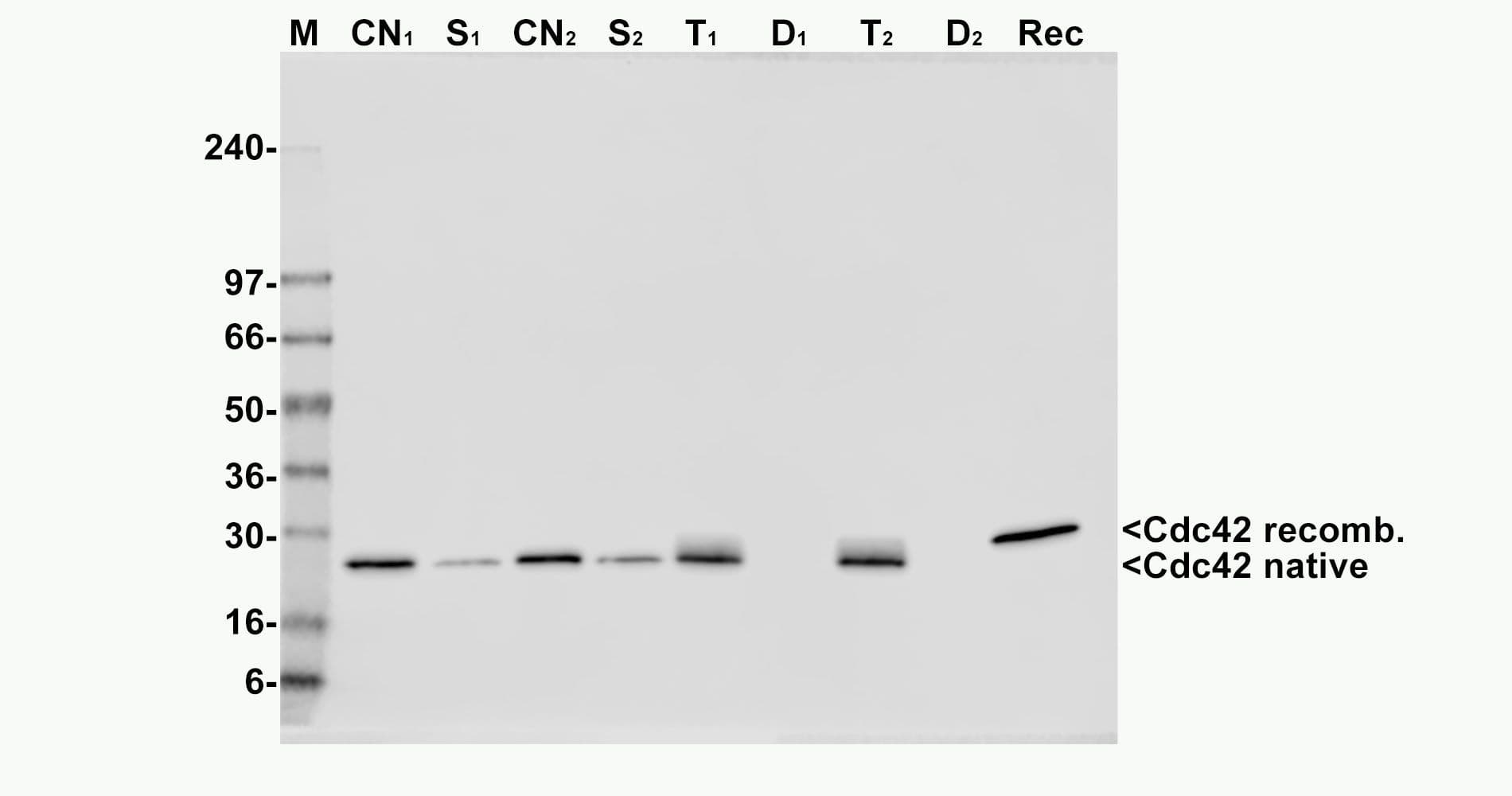
The Cdc42 activation assay was tested by loading the Cdc42 protein in cell lysates with either GTPγS or GDP. As expected, the GTPγS-loaded Cdc42 is very efficiently precipitated while very little GDP-loaded Cdc42 is precipitated (Fig. 2).

bk034fig2

Figure 2. Results from BK034 Cdc42 activation assay. Activated Cdc42 was precipitated and detected in a Western blot using kit BK034. The first lane shows a 50 ng recombinant His-tagged Cdc42 standard (Rec. His-Cdc42). The following lanes shows the pull-down of inactive, GDP-loaded Cdc42 (Cdc42-GDP PD) or active, GTPγS-loaded Cdc42 (Cdc42-GTP PD) from equal amounts of cell lysates.

For product Datasheets and MSDSs please click on the PDF links below.   For additional information, click on the FAQs tab above or contact our Technical Support department at tservice@cytoskeleton.com

AuthorTitleJournalYearArticle Link
Bai, Xiaoyuan et al.The extracellular cyclophilin A-integrin β2 complex as a therapeutic target of viral pneumoniaMolecular Therapy2024
Arnott, Malcolm et al.Transcription of microRNAs is regulated by developmental signaling pathways and transcription factorsFrontiers in Cell and Developmental Biology2024
Ha, Cuong P. et al.Humanin activates integrin αV–TGFβ axis and leads to glioblastoma progressionCell Death & Disease2024
Laurent-Issartel, Carine et al.Ascites microenvironment conditions the peritoneal pre-metastatic niche to promote the implantation of ovarian tumor spheroids: Involvement of fibrinogen/fibrin and αV and α5β1 integrinsExperimental Cell Research2024
Guo, Xiong et al.Rabenosyn-5 suppresses non-small cell lung cancer metastasis via inhibiting CDC42 activityCancer Gene Therapy 2024
Xu, Nana et al.CircCDC42-encoded CDC42-165aa regulates macrophage pyroptosis in Klebsiella pneumoniae infection through Pyrin inflammasome activationNature Communications2024
Yang, Ruicheng et al.Egr-1 is a key regulator of the blood-brain barrier damage induced by meningitic Escherichia coliCell Communication and Signaling2024
Veloso, Alexandra et al.The cytoskeleton adaptor protein Sorbs1 controls the development of lymphatic and venous vessels in zebrafishBMC Biology2024
Hu, Xinrong et al.A Natural Small Molecule Mitigates Kidney Fibrosis by Targeting Cdc42-mediated GSK-3β/β-catenin SignalingAdvanced Science2024
Newman, Daniel et al.3D matrix adhesion feedback controls nuclear force coupling to drive invasive cell migrationCell reports2023
Fang, Huan et al.Integrin β4 promotes DNA damage drug resistance in triple-negative breast cancer via TNFAIP2/IQGAP1/Rac1eLife2023
Yang, Yuchen et al.β-hydroxybutyrate impairs the directionality of migrating neutrophils through inhibiting the autophagy-dependent degradation of Cdc42 and Rac1 in ketotic cowsJournal of Dairy Science2023
Serwe, Guillaume et al.CNK2 promotes cancer cell motility by mediating ARF6 activation downstream of AXL signallingNature Communications2023
Spel, Lotte et al.CDC42 regulates PYRIN inflammasome assemblyCell Reports2022
Khan, Alamzeb et al.ArhGEF12 activates Rap1A and not RhoA in human dermal microvascular endothelial cells to reduce tumor necrosis factor-induced leakFASEB journal2022
Li, Xiaopeng et al.Hhex inhibits cell migration via regulating RHOA/CDC42-CFL1 axis in human lung cancer cellsCell Communication and Signaling2021
Li, Chunsen et al.GEFT Inhibits Autophagy and Apoptosis in Rhabdomyosarcoma via Activation of the Rac1/Cdc42-mTOR Signaling PathwayFrontiers in Oncology2021
Zhou, Yongjie et al.Congenital biliary atresia is correlated with disrupted cell junctions and polarity caused by Cdc42 insufficiency in the liverTheranostics2021
McCray, Brett A. et al.Neuropathy-causing TRPV4 mutations disrupt TRPV4-RhoA interactions and impair neurite extensionNature Communications2021
Chen, Lixia et al.CSRP2 suppresses colorectal cancer progression via p130Cas/Rac1 axis-meditated ERK, PAK, and HIPPO signaling pathwaysTheranostics2020
Lian, Eric Y. et al.RET isoforms contribute differentially to invasive processes in pancreatic ductal adenocarcinomaOncogene2020
Zhang, Jiawei et al.In vivo and in vitro activation of dormant primordial follicles by EGF treatment in mouse and humanClinical and Translational Medicine2020
Hosseini, Kamran et al.EMT-Induced Cell-Mechanical Changes Enhance Mitotic Rounding StrengthAdvanced Science2020
Gu, Jiawen et al.Rho-GEF trio regulates osteoclast differentiation and function by Rac1/Cdc42Experimental Cell Research2020
Lang, Yue et al.MiR-30 family prevents uPAR-ITGB3 signaling activation through calcineurin-NFATC pathway to protect podocytesCell Death and Disease2019
Carvalho, J. R. et al.Non-canonical Wnt signaling regulates junctional mechanocoupling during angiogenic collective cell migrationeLife2019
Dagliyan, Onur et al.Engineering proteins for allosteric control by light or ligandsNature Protocols2019
Zhou, Yi Fan et al.Sema3E/PlexinD1 signaling inhibits postischemic angiogenesis by regulating endothelial DLL4 and filopodia formation in a rat model of ischemic strokeFASEB Journal2019
Liu, Chunxia et al.Epigenetically upregulated GEFT-derived invasion and metastasis of rhabdomyosarcoma via epithelial mesenchymal transition promoted by the Rac1/Cdc42-PAK signalling pathwayEBioMedicine2019
Yang, Huan et al.Cytotoxic Necrotizing Factor 1 Downregulates CD36 Transcription in Macrophages to Induce Inflammation During Acute Urinary Tract InfectionsFrontiers in Immunology2018
Veluthakal, Rajakrishnan et al.Restoration of glucose-stimulated Cdc42-PAK1 activation and insulin secretion by a selective Epac activator in type 2 diabetic human isletsDiabetes2018
Sepúlveda-Ramírez, Silvia P. et al.Cdc42 controls primary mesenchyme cell morphogenesis in the sea urchin embryoDevelopmental Biology2018
Chavali, Manideep et al.Non-canonical Wnt signaling regulates neural stem cell quiescence during homeostasis and after demyelinationNature Communications2018
Sunkavalli, Ushasree et al.Analysis of host microRNA function uncovers a role for miR-29b-2-5p in Shigella capture by filopodiaPLoS Pathogens2017
Vidal-Quadras, Maite et al.Endocytic turnover of Rab8 controls cell polarizationJournal of Cell Science2017
Yao, Zhihui et al.P311 Accelerates Skin Wound Reepithelialization by Promoting Epidermal Stem Cell Migration Through RhoA and Rac1 ActivationStem Cells and Development2017
Guo, Yaxiu et al.Cytotoxic necrotizing factor 1 promotes prostate cancer progression through activating the Cdc42–PAK1 axisJournal of Pathology2017
Tormos, Ana M. et al.P38α regulates actin cytoskeleton and cytokinesis in hepatocytes during development and agingPLoS ONE2017
Saito, Masaki et al.Tctex‐1 controls ciliary resorption by regulating branched actin polymerization and endocytosisEMBO reports2017
Jia, L. et al.KLF5 promotes breast cancer proliferation, migration and invasion in part by upregulating the transcription of TNFAIP2Oncogene2016
Zhan, Rixing et al.Nitric oxide promotes epidermal stem cell migration via cGMP-Rho GTPase signallingScientific Reports2016
Moodley, Serisha et al.Stimulus-dependent dissociation between XB130 and Tks5 scaffold proteins promotes airway epithelial cell migrationOncotarget2016
Zhan, Rixing et al.Nitric oxide enhances keratinocyte cell migration by regulating Rho GTPase via cGMP-PKG signallingPLoS ONE2015
Ricotti, Leonardo et al.Boron nitride nanotube-mediated stimulation modulates F/G-actin ratio and mechanical properties of human dermal fibroblastsJournal of Nanoparticle Research2014
Tang, Xiaoyun et al.Lipid phosphate phosphatase-1 expression in cancer cells attenuates tumor growth and metastasis in miceJournal of Lipid Research2014
Knowles, Byron C. et al.Myosin Vb uncoupling from RAB8A and RAB11A elicits microvillus inclusion diseaseJournal of Clinical Investigation2014
Nithipatikom, Kasem et al.Cannabinoid receptor type 1 (CB1) activation inhibits small GTPase RhoA activity and regulates motility of prostate carcinoma cellsEndocrinology2012
Sakabe, Isamu et al.Age-related guanine nucleotide exchange factor, mouse Zizimin2, induces filopodia in bone marrow-derived dendritic cellsImmunity and Ageing2012
Asai, Akira et al.Involvement of Rac GTPase activation in phosphatidylcholine hydroperoxide-induced THP-1 cell adhesion to ICAM-1Biochemical and biophysical research communications2011
Nikolic, Damjan S. et al.HIV-1 activates Cdc42 and induces membrane extensions in immature dendritic cells to facilitate cell-to-cell virus propagationBlood2011
Broman, Michael T. et al.Cdc42 regulates adherens junction stability and endothelial permeability by inducing α-catenin interaction with the vascular endothelial cadherin complexCirculation Research2006
Slice, Lee W. et al.Angiotensin II and epidermal growth factor induce cyclooxygenase-2 expression in intestinal epithelial cells through small GTPases using distinct signaling pathwaysThe Journal of biological chemistry2005
Nur-E-Kamal, Alam et al.Three dimensional nanofibrillar surfaces induce activation of RacBiochemical and biophysical research communications2005
Tang, Dale D. et al.The adapter protein CrkII regulates neuronal Wiskott-Aldrich syndrome protein, actin polymerization, and tension development during contractile stimulation of smooth muscleJournal of Biological Chemistry2005
Chellaiah, Meenakshi A.Regulation of actin ring formation by Rho GTPases in osteoclastsJournal of Biological Chemistry2005
Sasai, Noriaki et al.The neurotrophin-receptor-related protein NRH1 is essential for convergent extension movementsNature Cell Biol2004
Tang, Dale D. et al.The small GTPase Cdc42 regulates actin polymerization and tension development during contractile stimulation of smooth muscleJournal of Biological Chemistry2004
Liu, Xiu-Fen et al.Nucleotide Exchange Factor ECT2 Interacts with the Polarity Protein Complex Par6/Par3/Protein Kinase Cζ (PKCζ) and Regulates PKCζ ActivityMolecular and Cellular Biology2004
Vogl, Thomas et al.MRP8 and MRP14 control microtubule reorganization during transendothelial migration of phagocytesBlood2004


Question 1: I have high background and/or multiple bands on my western blot.  How can I fix this?

Answer 1:  There are multiple causes of high background and/or multiple bands.  Some suggestions to improve background signal include:

  1.  When blotting use 70v for 45min only as the small G-proteins are very mobile.
  2. Fully remove SDS from the gel by using a non-SDS containing buffer for transfer and performing a full 15 min gel wash step in the transfer buffer before blotting.
  3. Dry the PVDF membrane for 30 min after transfer and before blocking (not necessary for nitrocellulose)
  4. Making sure that the TBST contains 10 mM Tris, 0.05% Tween 20 and 150 mM NaCl.
  5. Incubating with the primary antibody overnight at 4°C and using the appropriate ECL detection system. 


Question 2: How much of the beads should I use for my pull-down experiments?

Answer 2:  PAK-PBD-GST beads (Cat. # PAK02) will bind to Cdc42-GDP with a much lower affinity than Cdc42-GTP.  If too many PAK-PBD beads are added to the pull-down assay, there will be significant binding to inactive (GDP-bound) Cdc42.  The result of this will be an underestimation of Cdc42 activation.  For this reason, we highly recommend performing a bead titration to determine optimal conditions for any given Cdc42 activation or inactivation assay.  Once optimal conditions have been established, bead titrations should no longer be necessary.  We recommend 10, 15 and 20 μg bead titrations.


Question 3:  How can I test whether the beads are working properly?

Answer 3:  A standard biological assay for PAK-PBD GST protein beads consists of a Cdc42 protein pull-down from cells loaded with either GTPγS (Cat. # BS01) or GDP.  Here are guidelines to follow (see Cat. # PAK02 or BK034 datasheets for more details):


Positive Cellular Protein Control:

Total cell lysate (300 – 800 μg) should be loaded with GTPγS as a positive control for the pull-down assay.  The following reaction details how to load endogenous Cdc42 with the nonhydrolysable GTP analog (GTPγS).  This is an excellent substrate for PAK-PBD beads and should result in a strong positive signal in a pull-down assay.


a) Perform GTP loading on 300 – 800 μg of cell lysate (0.5 mg/ml protein concentration) by adding 1/10th volume of Loading Buffer.

b) Immediately add 1/100th volume of GTPγS (200 μM final concentration). Under these conditions, 5 - 10% of the Cdc42 protein will load with non-hydrolysable GTPγS and will be “pulled-down” with the PAK-PBD beads in the assay.

c) Incubate the control sample at 30°C for 15 min with gentle rotation.

d) Stop the reaction by transferring the tube to 4°C and adding 1/10th volume of STOP Buffer.

e) Use this sample immediately in a pull-down assay.


Negative Cellular Protein Control:

This reaction should be performed in an identical manner to the Positive Control reaction except that 1/100th volume of GDP (1 mM final concentration) should be added to the reaction in place of the GTPγS.  Loading endogenous Cdc42 with GDP will inactivate Cdc42 and this complex will bind very poorly to PAK-PBD beads.


If you have any questions concerning this product, please contact our Technical Service department at tservice@cytoskeleton.com