Fibronectin (Fluorescent Red/Orange HiLyte Fluor™ 555 Labeled)
Background Information
The Extracellular Matrix (ECM) is composed of collagen, non-collagenous glycoproteins and proteoglycans. These components are secreted from cells to create an ECM meshwork that surrounds cells and tissues. The ECM regulates many aspects of cellular function, including the cells dynamic behavior, cytoskeletal organization, and intercellular communication (1).
Fibronectin is a high-molecular weight (~440kDa) glycoprotein found in the extracellular matrix and blood plasma. It is made up of two subunits that vary in size between 235 and 270 kDa (due to alternate splicing). The secreted fibronectin dimer is a soluble protein which polymerizes to higher order fibrils in the ECM.
Fibronectin plays a major role in cell adhesion, growth, migration, actin dynamics and differentiation, and it is important for processes such as wound healing and embryonic development (2). These functions are mediated through fibronectin binding to integrin receptor proteins (2). Altered fibronectin expression, degradation, and organization have been associated with a number of pathologies, including cancer and fibrosis (3).
In addition to integrins, fibronectin binds extracellular matrix components such as collagen, fibrin, and heparan sulfate proteoglycans (e.g. syndecans).
Product Uses
Material
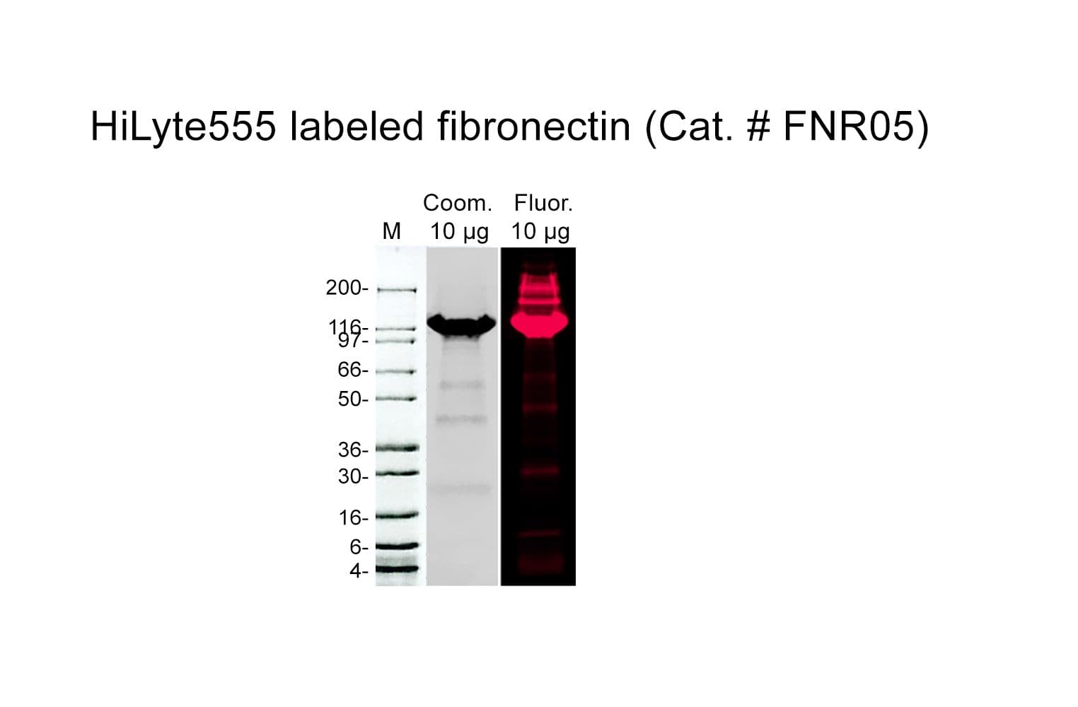
Fibronectin is purified from bovine plasma. Protein purity is determined by scanning densitometry of Coomassie Blue stained protein on a 4-20% polyacrylamide gel. Red fluorescent fibronectin is >80% pure (Figure 1).
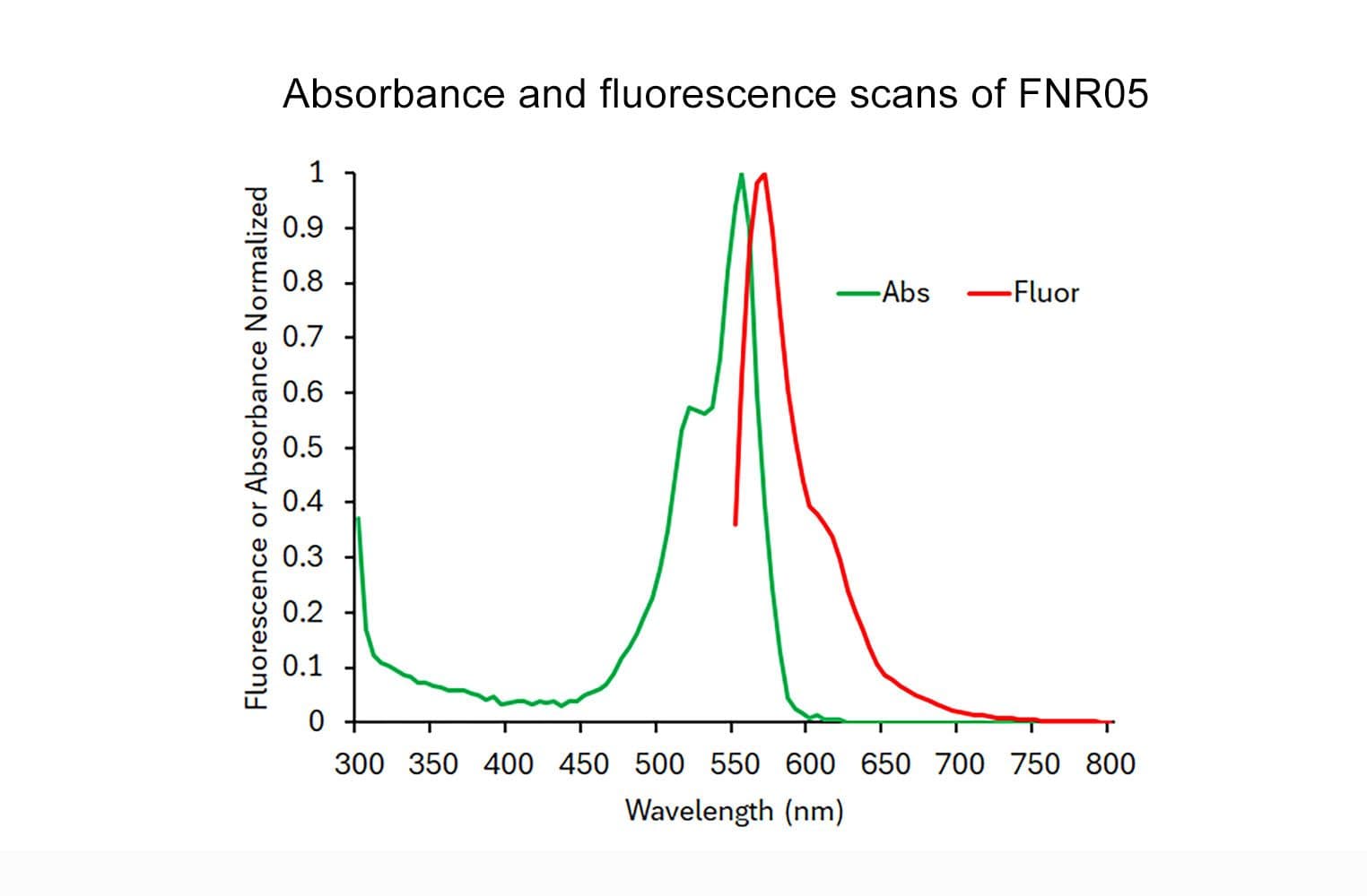
The protein is modified to contain covalently linked red/orange fluorescent HiLyte Fluor™ 555 at random surface lysines. An activated ester of HiLyte Fluor™ 555 [succinimidyl ester] is used to label the protein. Labeling stoichiometry is determined by spectroscopic measurement of protein and dye concentrations. The final labeling stoichiometry is 1-3 dyes per protein molecule (Figure 2). No free dye is apparent in the final product (Figure 1). Red fluorescent fibronectin can be detected using a filter set of 540nm excitation and 590 nm emission, or laser 561nm excitation and 590nm emission filter.
Fibronectin runs as individual subunits on SDS-PAGE with an apparent molecular weight of 220 kDa. FNR05 is supplied as a pink/red lyophilized powder. Each vial of FNR05 contains 20 µg protein.
HiLyte Fluor™ is a trademark of Anaspec Inc (CA, USA)
Purity
Figure 1: Red/Orange fluorescent fibronectin Purity Determination
Legend: 20 µg of FNR05 was analyzed by electrophoresis in a 4-20% SDS-PAGE system. A LI-COR Odessy gel analysis was performed at 700nm (Coomassie, lane 1), and 600nm (HiLyte555, lane 2). The Red fluorescent labeled protein was visualized at 230 kDal and no free dye was observed in the dye front. Protein quantitation was determined with the Precision Red™ Protein Assay Reagent (Cat. # ADV02). Mark12 molecular weight markers are from Invitrogen.
Figure 2: Absorbance and fluorescence scan of Red/Orange fluorescent fibronectin in solution
Legend: FNR05 was diluted with nanopure water and its absorbance (blue line) and fluorescence (red line, with emission 570 nm) spectra were scanned between 350 and 850 nm. Red fluorescent labeling stoichiometry was calculated to be 1-3 dyes per fibronectin protein using the absorbance maximum for Red fluorescence at 555 nm and the Beer-Lambert law. The extinction coefficient of the dye is 150,000 M-1cm-1.
Storage and Reconstitution
Shipped at ambient temperature. The lyophilized protein can be stored desiccated to <10% humidity at 4°C for 6 months in the dark. For reconstitution, briefly centrifuge to collect the product at the bottom of the tube and resuspend to 1 mg/ml with 20 µl room temperature sterile nanopure water. Let the protein re-dissolve for 1-2 minutes without mixing; after 1-2 minutes the protein solution can be gently pipetted up and down 2-3 times to ensure complete resuspension. Excessive mixing should be avoided as this can cause protein aggregation. Once the product is resuspended, place it on ice. The protein will be in the following buffer: 20 mM Tris-HCl pH 7.6, 20 mM NaCl, 0.1 mM EDTA, 15 mM BME, and 5% (w/v) sucrose. The concentrated protein can be aliquoted into experiment sized amounts, snap frozen in liquid nitrogen and stored at -70°C where it is stable for 6 months. For working concentrations, further dilution of Red/Orange fluorescent fibronectin 555 should be made in a suitable buffer or tissue culture media. Fluorescent fibronectin is a labile protein and should be handled with care. Avoid repeated freeze-thaw cycles.
Biological Activity Assay
Proteolytic degradation of the ECM is a critical step during cell invasion and is necessary for both physiological and pathological processes. Red fluorescent fibronectin can be used as an ECM substrate to monitor invasion through observation of ECM degradation (4).
References
Product Citations / Related Products
For the latest citations and related products please visit www.cytoskeleton.com
For product Datasheets and MSDSs please click on the PDF links below. For additional information, click on the FAQs tab above or contact our Technical Support department at tservice@cytoskeleton.com
If you have any questions concerning this product, please contact our Technical Service department at tservice@cytoskeleton.com