Ras Pull-down Activation Assay Biochem Kit (bead pull-down format) - 50 Assays

Ras Activation Assay Biochem Kit (bead pull-down format) - 50 Assays
$0.00

Product Uses Include

  • Analysis of in vivo Ras activation.

Introduction
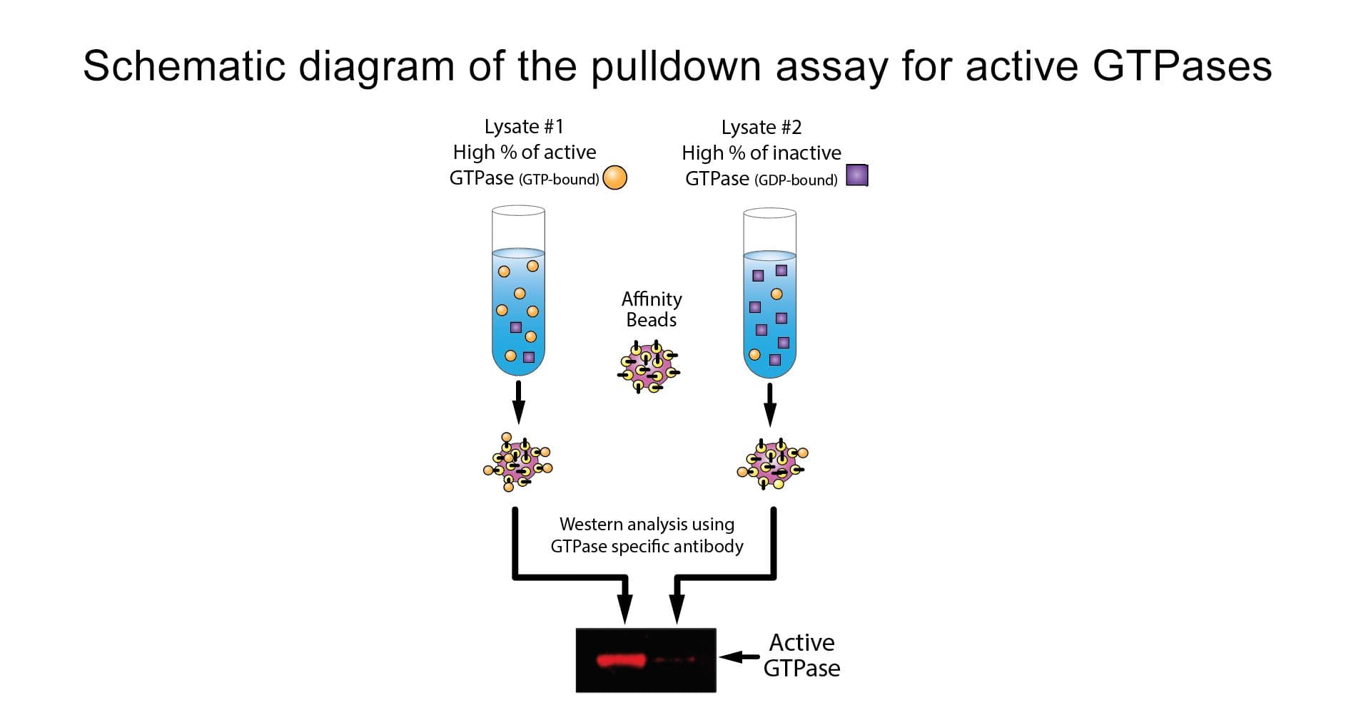
The Ras switch operates by alternating between an active, GTP-bound state and an inactive, GDP-bound state.  Understanding the mechanisms that regulate activation / inactivation of Ras-like GTPases is of obvious biological significance and is a subject of intense investigation.  The fact that Ras family effector proteins will specifically recognize the GTP bound form of the protein has been exploited experimentally to develop a powerful affinity purification assay that monitors Ras protein activation.  The assay uses the Ras-binding domain (RBD) of the Ras effector kinase Raf1.  The Raf-RBD domain has been shown to bind specifically to the GTP-bound form of Ras proteins.  The fact that the Raf-RBD has a high affinity for GTP-Ras and that its binding results in a significantly reduced intrinsic and catalytic rate of hydrolysis of Ras make it an ideal tool for affinity purification of GTP-Ras from cell lysates.  The Raf-RBD is in the form of a GST fusion protein, which allows one to "pull-down" the Raf-RBD/GTP-Ras complex with glutathione affinity beads.  The assay therefore provides a simple means of quantitating Ras activation in cells.  The amount of activated Ras is determined by a western blot using a Ras specific antibody.

Kit contents
The kit contains enough materials for 50 assays, depending on assay setup, and includes reagents for positive and negative controls. The following components are included:

  1. GST tagged Raf1-RBD protein bound to colored glutathione agarose beads (Cat. # RF02).
  2. p21 Ras monoclonal antibody
  3. Cell lysis Buffer
  4. Protease inhibitor cocktail (Cat. # PIC02)
  5. Wash Buffer
  6. Loading Buffer
  7. STOP Buffer
  8. GTPγS (non-hydrolyzable GTP analog, Cat. # BS01)
  9. GDP
  10. Manual with detailed protocols and extensive troubleshooting guide.
beads

 Figure 1. The brightly colored glutathione agarose beads in kit BK008 makes the kit easy to use..

Equipment needed

  1. SDS-PAGE minigel system and western blotting transfer apparatus

Associated Products:

G-LISA Products:
Rac1 G-LISA™ Activation Assay, luminescence format (Cat.# BK126)
Rac1,2,3 G-LISA™ Activation Assay, colorimetric format (Cat.# BK125)
RhoA G-LISA™ Activation Assay, colorimetric format (Cat.# BK124)
RhoA G-LISA™ Activation Assay, luminescence format (Cat.# BK121)
Cdc42 G-LISA™ Activation Assay, colorimetric format (Cat.# BK127)

Associated Products:
Anti-Cdc42 monoclonal antibody (Cat.# ACD03)
Anti-Rac1 monoclonal antibody (Cat.# ARC03)
Anti-RhoA monoclonal antibody (Cat.# ARH04)

For product Datasheets and MSDSs please click on the PDF links below.   For additional information, click on the FAQs tab above or contact our Technical Support department at tservice@cytoskeleton.com

AuthorTitleJournalYearArticle Link
Han, David et al.The RAL Small G Proteins Are Clinically Relevant Targets in Triple Negative Breast CancerCancers2024
Wang, Xuan et al.Triple Blockade of Oncogenic RAS Signaling Using KRAS and MEK Inhibitors in Combination with Irradiation in Pancreatic CancerInternational Journal of Molecular Sciences2024
Consalvo, Kristen M. et al.PTEN and the PTEN-like phosphatase CnrN have both distinct and overlapping roles in a Dictyostelium chemorepulsion pathwayJournal of Cell Science2024
Carrión-Estrada, Dayan A. et al.Antineoplastic effect of compounds C14 and P8 on TNBC and radioresistant TNBC cells by stabilizing the K-Ras4BG13D/PDE6δ complexFrontiers in Oncology2024
Najumudeen, Arafath K. et al.KRAS allelic imbalance drives tumour initiation yet suppresses metastasis in colorectal cancer in vivoNature Communications2024
Ravishankar, Rajani et al.Specific Disruption of Ras2 CAAX Proteolysis Alters Its Localization and FunctionMicrobiology Spectrum2023
Rubio, Karla et al.Non-canonical integrin signaling activates EGFR and RAS-MAPK-ERK signaling in small cell lung cancerTheranostics2023
Prahallad, Anirudh et al.CRISPR Screening Identifies Mechanisms of Resistance to KRASG12C and SHP2 Inhibitor Combinations in Non-Small Cell Lung CancerCancer research2023
Serwe, Guillaume et al.CNK2 promotes cancer cell motility by mediating ARF6 activation downstream of AXL signallingNature Communications2023
Bortolami, Alessandro et al.Integrin-KCNB1 potassium channel complexes regulate neocortical neuronal development and are implicated in epilepsyCell Death & Differentiation2022
Cruz-Nova, Pedro et al.131I-C19 Iodide Radioisotope and Synthetic I-C19 Compounds as K-Ras4B–PDE6δ Inhibitors: A Novel Approach against Colorectal Cancer—Biological Characterization, Biokinetics and DosimetryMolecules2022
Zhu, Dantong et al.Loss of PTEN-Induced Kinase 1 Regulates Oncogenic Ras-Driven Tumor Growth By Inhibiting Mitochondrial FissionFrontiers in Oncology2022
Carnevale, Julia et al.RASA2 ablation in T cells boosts antigen sensitivity and long-term functionNature2022
Jeon, So Mi et al.Blockade of PD-L1/PD-1 signaling promotes osteo-/odontogenic differentiation through Ras activationInternational Journal of Oral Science2022
Song, Dan et al.NSUN2-mediated mRNA m5C Modification Regulates the Progression of Hepatocellular CarcinomaGenomics, Proteomics & Bioinformatics2022
Kirolos, Sara A. et al.A chemorepellent inhibits local Ras activation to inhibit pseudopod formation to bias cell movement away from the chemorepellentMolecular biology of the cell2022
Ablain, Julien et al.SPRED1 deletion confers resistance to MAPK inhibition in melanomaThe Journal of experimental medicine2021
Tulpule, Asmin et al.Kinase-mediated RAS signaling via membraneless cytoplasmic protein granulesCell2021
Wiley, Christopher D. et al.Oxylipin biosynthesis reinforces cellular senescence and allows detection of senolysisCell Metabolism2021
Yan, Wupeng et al.Structural Insights into the SPRED1-Neurofibromin-KRAS Complex and Disruption of SPRED1-Neurofibromin Interaction by Oncogenic EGFRCell Reports2020
Brandt, Raphael et al.Cell type-dependent differential activation of ERK by oncogenic KRAS in colon cancer and intestinal epitheliumNature Communications2019
Hood, Fiona E. et al.Isoform-specific Ras signaling is growth factor dependentMolecular Biology of the Cell2019
Rijal, Ramesh et al.An endogenous chemorepellent directs cell movement by inhibiting pseudopods at one side of cellsMolecular Biology of the Cell2019
Cai, Diana et al.Identification and characterization of oncogenic SOS1 mutations in lung adenocarcinomaMolecular Cancer Research2019
Pilling, Darrell et al.Different Isoforms of the Neuronal Guidance Molecule Slit2 Directly Cause Chemoattraction or Chemorepulsion of Human NeutrophilsThe Journal of Immunology2019
Cocco, Emiliano et al.Resistance to TRK inhibition mediated by convergent MAPK pathway activationNature Medicine2019
Dardaei, Leila et al.SHP2 inhibition restores sensitivity in ALK-rearranged non-small-cell lung cancer resistant to ALK inhibitorsNature Medicine2018
Saikia, Minakshi et al.Heteronemin, a marine natural product, sensitizes acute myeloid leukemia cells towards cytar***** chemotherapy by regulating farnesylation of RasOncotarget2018
Smolkin, Tatyana et al.Complexes of plexin-A4 and plexin-D1 convey semaphorin-3C signals to induce cytoskeletal collapse in the absence of neuropilinsJournal of Cell Science2018
Whiteside, Theresa L.Therapeutic targeting of oncogenic KRAS in pancreatic cancer by engineered exosomesTranslational Cancer Research2017
Malchers, Florian et al.Mechanisms of primary drug resistance in FGFR1-amplified lung cancerClinical Cancer Research2017
Lu, Xinyuan et al.MET exon 14 mutation encodes an actionable therapeutic target in lung adenocarcinomaCancer Research2017
Walton, Josephine B. et al.CRISPR/Cas9-derived models of ovarian high grade serous carcinoma targeting Brca1, Pten and Nf1, and correlation with platinum sensitivityScientific Reports2017
Ilinskaya, Olga N. et al.Direct inhibition of oncogenic KRAS by Bacillus pumilus ribonuclease (binase)Biochimica et Biophysica Acta - Molecular Cell Research2016
Larribere, Lionel et al.NF1 loss induces senescence during human melanocyte differentiation in an iPSC-based modelPigment Cell and Melanoma Research2015
Hrustanovic, Gorjan et al.RAS-MAPK dependence underlies a rational polytherapy strategy in EML4-ALK-positive lung cancerNature Medicine2015
Yan, Chao et al.Discovery and characterization of small molecules that target the GTPase RalNature2014
Du, Chang Qing et al.Inhibition of farnesyl pyrophosphate synthase prevents norep*********- induced fibrotic responses in vascular smooth muscle cells from spontaneously hypertensive ratsHypertension Research2014
Ciaglia, Elena et al.N6-isopentenyladenosine, an endogenous isoprenoid end product, directly affects cytotoxic and regulatory functions of human NK cells through FDPS modulationJournal of Leukocyte Biology2013
Kern, F. et al.Essential, non-redundant roles of B-Raf and Raf-1 in Ras-driven skin tumorigenesisOncogene2012
Stoppa, Giovanna et al.Ras signaling contributes to survival of human T-cell leukemia/lymphoma virus type 1 (HTLV-1) Tax-positive T-cellsApoptosis : an international journal on programmed cell death2012
Jiang, Xiao Sheng et al.Activation of Rho GTPases in Smith-Lemli-Opitz syndrome: pathophysiological and clinical implicationsHuman molecular genetics2010
Kowluru, Renu A.Role of Matrix Metalloproteinase-9 in the Development of Diabetic Retinopathy and Its Regulation by H-RasInvestigative Ophthalmology & Visual Science2010
Lito, Piro et al.Evidence that sprouty 2 is necessary for sarcoma formation by H-Ras oncogene-transformed human fibroblastsThe Journal of biological chemistry2008
Wasylyk, Christine et al.Inhibition of the Ras-Net (Elk-3) pathway by a novel pyrazole that affects microtubulesCancer Research2008


Question 1: I have high background and/or multiple bands on my western blot.  How can I fix this?

Answer 1:  There are multiple causes of high background and/or multiple bands.  Some suggestions to improve background signal include:

  1. When blotting use 70v for 45min only as the small G-proteins are very mobile.
  2. Fully remove SDS from the gel by using a non-SDS containing buffer for transfer and performing a full 15 min gel wash step in the transfer buffer before blotting.
  3. Dry the PVDF membrane for 30 min after transfer and before blocking (not necessary for nitrocellulose)
  4. Making sure that the TBST contains 10 mM Tris, 0.05% Tween 20 and 150 mM NaCl.
  5. Incubating with the primary antibody overnight at 4°C and using the appropriate ECL detection system. 


Question 2: How much of the beads should I use for my pull-down experiments?

Answer 2:  Raf-RBD beads (Cat. # RF02) will bind to Ras-GDP with a much lower affinity than Ras-GTP.  If too many Raf-RBD beads are added to the pull-down assay there will be significant binding to inactive (GDP-bound) Ras. The result of this will be an underestimation of Ras activation.  For this reason, we highly recommend performing a bead titration to determine optimal conditions for any given Ras activation or inactivation assay.  Once optimal conditions have been established, bead titrations should no longer be necessary. We recommend 20, 40 and 60 μl (66, 132 and 198 µg) bead titrations.


Question 3:  How can I test whether the beads are working properly?

Answer 3:  A standard biological assay for Raf-RBD beads consists of a Ras protein pull-down from cells loaded with either GTPγS (Cat. # BS01) or GDP.  Here are guidelines to follow (see Cat. # RF02 or BK008 datasheets for more details):


Positive Cellular Protein Control:

Total cell lysate (200 – 500 µg) should be loaded with GTPγS as a positive control for the pull-down assay. The following reaction details how to load endogenous Ras with the nonhydrolysable GTP analog (GTPγS).  This is an excellent substrate for Raf-RBD beads and should result in a strong positive signal in a pull-down assay.

a)  Perform GTP loading on 200 – 500 μg of cell lysate that is at a protein concentration between 0.4 – 2.0 mg/ml  by adding 1/10th volume of Loading Buffer.

b)  Immediately add 1/100th volume of GTPγS (200 μM final concentration). Under these conditions, 5 - 10% of the Ras protein will load with non-hydrolysable GTPγS and will be “pulled-down” with the Raf-RBD beads in the assay.

c)  Incubate the control sample at 37°C for 30 minutes with gentle rotation.

d)  Stop the reaction by transferring the tube to 4°C and adding 1/10th volume of STOP Buffer.

e)  Use this sample in a pull-down assay immediately.


Negative Cellular Protein Control:

This reaction should be performed in an identical manner to the Positive Control reaction except that 1/100th volume of GDP (1 mM final concentration) should be added to the reaction in place of the GTPγS. Loading endogenous Ras with GDP will inactivate Ras and this will bind very poorly to Raf-RBD beads.


If you have any questions concerning this product, please contact our Technical Service department at tservice@cytoskeleton.com