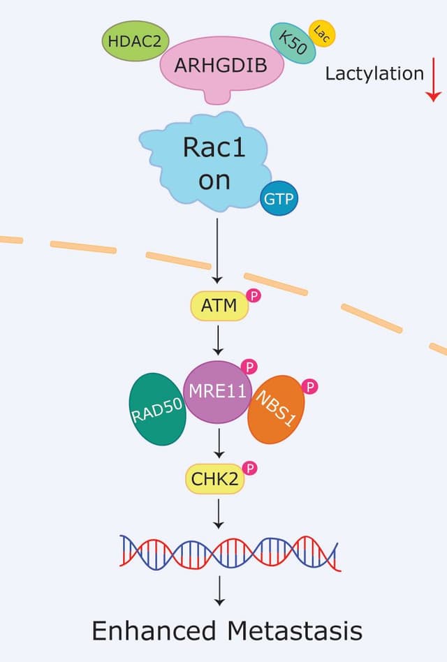
Can calcium signaling modulate MPS dynamic changes?
A critical property of actin is its dynamicity, which allows for the rapid production or destruction of actin structures to control cellular processes. This dynamic ability of actin is particularly important in neurons, which have both long-term, highly stable cellular structures and active remodeling structures. As discussed above, the MPS is primarily thought to provide mechanical support to the axon, implying that it may be a very stable actin structure. Conversely, the MPS studies on neurodegeneration and RTK signaling discussed above suggest that it can disassemble in response to stimuli, thus demonstrating dynamic properties. A recent study by Heller et al. showed that the MPS is actually highly dynamic and is constitutively remodeled by calcium signaling in neuronal cells23. Using lattice structured illumination microscopy (SIM), the group found that MPS remodeling driven by calcium signaling resulted in spectrin degradation by calpain and PKC-mediated adducin phosphorylation23. This is quite distinct from another recent study that showed that F-actin remodeling is highly active during the early development of the axonal MPS, but is less active in mature MPS (preprint). While there is still much to understand about MPS dynamics, it does appear that it has the capacity to undergo dynamic changes in response to multiple different types of stimuli.
Summary and future directions of discovery
Detection of these specialized MPS structures has boosted our understanding of the role that actin plays in the axons of neurons to regulate its structure, receptor signaling, and overall health of these specialized cells. Moreover, several other functions of the MPS have also been identified, including organizing membrane proteins, mechanosensation, neuronal excitability, microtubule crosstalk, and ion channel distribution(reviewed in 9, 19). Its importance in neurobiology is further supported by reports showing that several neurological diseases are linked to hereditary mutations of key MPS component proteins24, and genetic disruption of the MPS in mice leads to neurological impairment25, 26.Collectively, it is clear that the MPS is a critical actin structure that has a profound effect on neuronal health and function and may someday be a potential target to treat neurodegenerative diseases.
References
1. Xu, K., G. Zhong, and X. Zhuang, Actin, spectrin, and associated proteins form a periodic cytoskeletal structure in axons. Science, 2013. 339(6118): p. 452-6.
2. Leterrier, C., Putting the axonal periodic scaffold in order. Curr Opin Neurobiol, 2021. 69: p. 33-40.
3. Qu, Y., et al., Periodic actin structures in neuronal axons are required to maintain microtubules. Mol Biol Cell, 2017. 28(2): p. 296-308.
4. Macarron-Palacios, V., et al., Paralemmin-1 controls the nanoarchitecture of the neuronal submembrane cytoskeleton. Sci Adv, 2025. 11(10): p. eadt3724.
5. D'Este, E., et al., Ultrastructural anatomy of nodes of Ranvier in the peripheral nervous system as revealed by STED microscopy. Proc Natl Acad Sci U S A, 2017. 114(2): p. E191-E199.
6. Zhou, R., et al., Proteomic and functional analyses of the periodic membrane skeleton in neurons. Nat Commun, 2022. 13(1): p. 3196.
7. He, J., et al., Prevalent presence of periodic actin-spectrin-based membrane skeleton in a broad range of neuronal cell types and animal species. Proc Natl Acad Sci U S A, 2016. 113(21): p. 6029-34.
8. Li, N., et al., Structural basis of membrane skeleton organization in red blood cells. Cell, 2023. 186(9): p. 1912-1929 e18.
9. Costa, A.R. and M.M. Sousa, The role of the membrane-associated periodic skeleton in axons. Cell Mol Life Sci, 2021. 78(13): p. 5371-5379.
10. Unsain, N., F.D. Stefani, and A. Caceres, The Actin/Spectrin Membrane-Associated Periodic Skeleton in Neurons. Front Synaptic Neurosci, 2018. 10: p. 10.
11. Wernert, F., et al., The actin-spectrin submembrane scaffold restricts endocytosis along proximal axons. Science, 2024. 385(6711): p. eado2032.
12. Lorenzo, D.N., et al., betaII-spectrin promotes mouse brain connectivity through stabilizing axonal plasma membranes and enabling axonal organelle transport. Proc Natl Acad Sci U S A, 2019. 116(31): p. 15686-15695.
13. Costa, A.R., et al., The membrane periodic skeleton is an actomyosin network that regulates axonal diameter and conduction. Elife, 2020. 9.
14. Wang, T., et al., Radial contractility of actomyosin rings facilitates axonal trafficking and structural stability. J Cell Biol, 2020. 219(5).
15. Berth, S.H. and T.E. Lloyd, Disruption of axonal transport in neurodegeneration. J Clin Invest, 2023. 133(11).
16. Hauser, M., et al., The Spectrin-Actin-Based Periodic Cytoskeleton as a Conserved Nanoscale Scaffold and Ruler of the Neural Stem Cell Lineage. Cell Rep, 2018. 24(6): p. 1512-1522.
17. Zhou, R., et al., Membrane-associated periodic skeleton is a signaling platform for RTK transactivation in neurons. Science, 2019. 365(6456): p. 929-934.
18. Zheng, Y., et al., A chemically inducible multimerization system for tunable and background-free RTK activation. bioRxiv, 2025.
19. Gallo, G., The Axonal Actin Filament Cytoskeleton: Structure, Function, and Relevance to Injury and Degeneration. Mol Neurobiol, 2024. 61(8): p. 5646-5664.
20. Morrow, J.S. and M.C. Stankewich, The Spread of Spectrin in Ataxia and Neurodegenerative Disease. J Exp Neurol, 2021. 2(3): p. 131-139.
21. Unsain, N., et al., Remodeling of the Actin/Spectrin Membrane-associated Periodic Skeleton, Growth Cone Collapse and F-Actin Decrease during Axonal Degeneration. Sci Rep, 2018. 8(1): p. 3007.
22. Wang, G., et al., Structural plasticity of actin-spectrin membrane skeleton and functional role of actin and spectrin in axon degeneration. Elife, 2019. 8.
23. Heller, E., N. Kurup, and X. Zhuang, The membrane skeleton is constitutively remodeled in neurons by calcium signaling. Science, 2025. 389(6760): p. eadn6712.
24. Li, S., et al., Spectrins and human diseases. Transl Res, 2022. 243: p. 78-88.
25. Huang, C.Y., et al., alphaII Spectrin Forms a Periodic Cytoskeleton at the Axon Initial Segment and Is Required for Nervous System Function. J Neurosci, 2017. 37(47): p. 11311-11322.
26. Huang, C.Y., et al., An alphaII Spectrin-Based Cytoskeleton Protects Large-Diameter Myelinated Axons from Degeneration. J Neurosci, 2017. 37(47): p. 11323-11334.