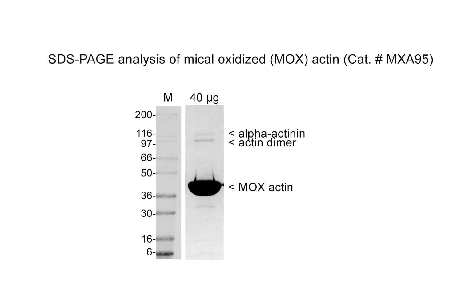
MICAL-Oxidized Actin Protein (>95% pure): Rabbit Skeletal Muscle

MICAL-Oxidized Actin Protein (>95% pure): Rabbit Skeletal Muscle (250 micrograms)
$0.00
SigSeekGraphic2

MICAL interacts with F-actin and uses NADPH as a cofactor to oxidize actin at Met44 and Met47 (b-actin nomenclature). Functionally, oxidation of Met44 has a profound effect on actin polymerization because the residue resides in the D-loop of subdomain 2 of the protein, which is critical for actin subunit contacts; thus, upon oxidation, Met44 becomes negatively charged and interferes with actin monomer-monomer interaction and promotes F-actin severing and depolymerization. 
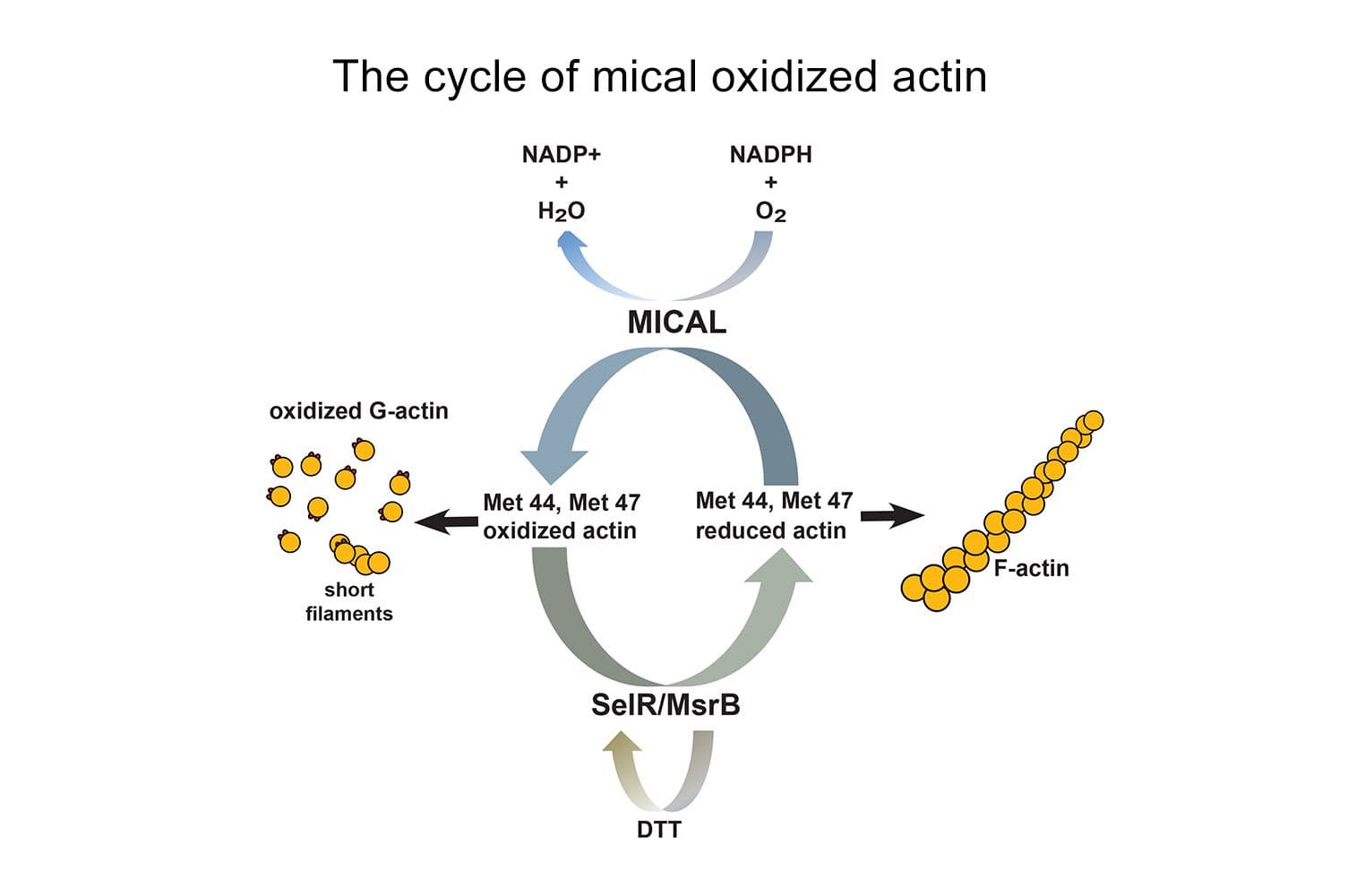
Regulation of actin oxidation at Met44/Met47 has been shown to destabilize F-actin in vivo and to play a key role in a growing number of cellular processes.  As part of the MOXtrue™ product line, rabbit skeletal muscle actin protein (MICAL-oxidized) (MXA95) has been enzymatically oxidized at methionines 44 and 47  with the MICAL flavoprotein monoxygenase protein. Purified MICAL-oxidized actin has reduced susceptibility to subtilisin A cleavage at M47/G48 by > 90%, and has also been validated for downstream applications such as sedimentation assays. 

To learn more about using MICAL-oxidized Actin as a research tool see our datasheet


To learn more about the MICAL/MsrB/Actin physiological redox system see our Newsletter 

Each lot of purified protein is quality controlled  to provide high batch to batch consistency, see COA documents.


Validated Applications

Actin Protein (MICAL-Oxidized) Purity Determination

A 50 μg sample of actin protein (MICAL-oxidized) was separated by electrophoresis in a 4- 20% tris-glycine gel and stained with Coomassie Blue. Protein quantitation was performed using the Precision RedTM Protein Assay Reagent (Cat. # ADV02). Mark12 standard molecular weight markers are from Invitrogen. 

MXA95_purity_gel_HQ

Subtilisin Assay on MICAL-Oxidized Actin vs Native Actin

Actin (Cat. # AKL99) and MICAL-oxidized actin (Cat. # MXA95) was diluted to 0.1 mg/ml (2.3 μM).  2 μg of each sample was then left untreated, or treated with subtilisin (1:200 w/w) for 15 min.  Samples were then separated by SDS-PAGE and visualized with Coomassie staining.  

Click here for a detailed method 

MXA95_subtilisin_gel_HQ

Actin Sedimentation Oxidized Versus Native Actin

Actin (Cat# AKL99) and MIcal-oxidized actin was diluted to 0.2 mg/ml (4.6 μM) or 0.8 mg/ml (18.4 μM) (see method). Samples were then incubated with 2x polymerization buffer at room temperature.  Samples were spun in an ultracentrifuge at 100,000 g for 1.5 h.  Samples were then separated by SDS-PAGE and visualized with Coomassie staining.

Click here for a detailed method 

MXA95_sedimentation_gel_HQ

For more information contact:  signalseeker@cytoskeleton.com

Associated Products:

MOXtrue™ 6xHis MICAL-1 Protein (Cat. # MIC01)

MOXtrue™ 6xHis MsrB2 Protein (Cat. # MB201)

MOXtrue™ MICAL-oxidized Pyrene Labeled Actin (Cat. # MPAX1)

Rabbit Skeletal Muscle Actin (Cat. # AKL95)

Pyrene Labeled Rabbit Skeletal Muscle Actin (Cat.# AP05)

For product Datasheets and MSDSs please click on the PDF links below.

Certificate of Analysis:  available upon request

If you have any questions concerning this product, please contact our Technical Service department at tservice@cytoskeleton.com

Visit our Signal-Seeker™ Tech Tips and FAQs page for technical tips and frequently asked questions regarding this and other Signal-Seeker™ products click here


If you have any questions concerning this product, please contact our Technical Service department at tservice@cytoskeleton.com