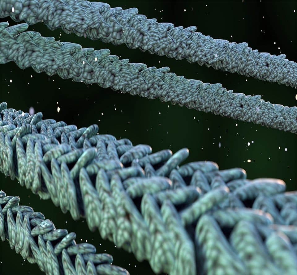
About Actin

Actin is a 43 kDa protein that is very highly conserved between species. Actin has three main isotypes (α-actin, β-actin and γ-actin), which show >90% amino-acid (aa) homology between isotypes and >98% homology within members of a particular isotypic group. The majority of the isotype heterogeneity is located in the amino-terminal 30 residues. The amino-terminus of actin is located on the periphery of the double-helix in F-actin (4) and this site is also thought to interact with myosin (5).


Actin isotypes have been shown to behave very differently in vitro and in vivo. Recent studies describe differential subcellular localization of γ-actin (1,2) and isotype specific binding of actin associated proteins. A good example of the latter phenomenon is the report of ezrin specificity for β-actin (3). These studies set a clear precedent that in vitro studies of actin systems should use the actin most closely related to the in vivo isotype.

Actin proteins, actin binding proteins, actin antibodies, actin buffers and research actin Biochem Kits™ are available for purchase.


G-actin polymerizes to form F-actin
Globular-actin (G-actin) readily polymerizes under physiological conditions to form Filamentous-actin (F-actin) with the concomitant hydrolysis of ATP. F-actin is a double-helical filament as shown in Figure 1


actin1


Figure 1. Double-helical structure of actin filaments.

Actin can polymerize from both ends in vitro. However, the rate of polymerization is not equal. This results in an intrinsic polarity in the actin filament. It has therefore become the convention to term the rapidly polymerizing end the plus-end (see Figure 1) or barbed-end while the slow growing end is called the minus-end or pointed-end.

The propensity of actin to polymerize is dependent upon the affinity of actin monomers for filament ends. Thus, there is an actin monomer concentration below which actin will not polymerize. This value has been termed the Critical Concentration (CC). At monomer concentrations above the CC, the actin will polymerize until the free monomer concentration is equal to the CC. When one is working with actin in vitro the extent of actin polymerization depends upon the conditions used. For example, at 4°C muscle actin has a CC of 0.03 mg/ml in the presence of 2 mM Mg2+ and 50 mM KCl, but when these ions are absent, the CC is greater than 3.0 mg/ml. Thus, by altering the ionic type and strength one can alter the amount of polymer formed. Non-muscle actin has its own CC values. For example, at 4°C in the presence of 2 mM Mg2+ and 50 mM KCl the CC is approximately 0.15 mg/ml. If Mg²+ and KCl are replaced with Ca²+, the CC will increase to nearly 10 mg/ml. Finally, the CC of non-muscle actin can be reduced to 0.03 mg/ml by increasing the temperature to 30°C (6).

Measuring actin polymer
Actin polymerization measurement is a quick and useful assay to evaluate the effect of a compound on actin function. The method of choice depends on the equipment available and the sensitivity required (see review ref. 7). The four most dependable methods are:

1) Fluorescence enhancement of pyrene conjugates
2) DNase inhibition assays
3) Viscosity measurements
4) Spin-down assays

1) By far the most versatile, sensitive and easiest to use is method 1. It involves mixing your control and test samples with a small amount of pyrene conjugated actin (see cat. # AP05) and then following polymerization with a fluorimeter. Fluorescence is enhanced up to twenty-fold by the association of the pyrene actin monomer into the polymer form (8 and Figure 2).


Actpol1


Figure 2. Polymerization of actin as measured by pyrene actin fluorescence.


Actin polymerization follows three phases. Just like in microtubule assembly, these are nuleation, growth and steady state as depicted in Figure 2. The extent of polymerization is indicated by the steady state level of flourescence, an upper limit can be measured by adding phalloidin (an actin stabilizing compound), which pushes the CC down to <0.01 mg/ml. This assay is greater than 95% accurate and the sample is not disturbed during the assay. Further details of setting up this assay are supplied with pyrene actin (AP05) and the Actin Polymerization Biochem Kit (BK003).

2) DNase inhibition assays are based on the high affinity interaction between G-actin and DNase 1, which results in inhibition of DNase activity (9). The value of this assay is that it can distinguish between G- and F-actin. In this regard it has been used to selectively assay the amounts of monomeric and filamentous actin in cell extracts. The assay has also been sucessfully adapted to study actin binding proteins that generate G-actin from actin filaments, an example of this is gelsolin protein (see Cat. # HPG6).

3) Viscosity measurements can be of the high or low shear variety. The choice depends on the specific aspect of polymerization that you wish to look at. For example, if you want to measure a small difference between short (1 µm) and medium (3-5 µm) actin filaments, then high shear methods is a good choice. For determining differences in samples with long filaments, on the other hand, a low shear apparatus is required. Apparatus for these experiments have been described by Pollard (10). Major problems with these methods for measuring actin assembly is that they disturb the reaction, i.e. shear the filaments, and they are not too accurate. However, viscosity measurements are good for comparing filament crosslinking between two samples (7).

4) Spin-down assays rely on differential sedimentation to separate F-actin from G-actin. Because of time constraints of the assay, the main use of this method is to obtain a quantitative value of actin polymer at steady state. This method can be used in combination with one of methods 1) or 2) to quantitate the amount of actin polymer over the time course of a reaction. This method has particular use for association studies (see later). This method is destructive, so can only be performed when all other measurements have been performed on the sample. A further potential drawback of this assay is that small actin oligos will not sediment, hence quantitative values may over-estimate the amount of actin monomer in the supernatant (11).

Uses of modified actins
Fluorescent actin can be used to study molecular dynamics in vivo (e.g. by microinjection into Xenopus eggs or cultured cells) and in vitro (e.g. actin treadmilling or myosin mobility assays)(12). It is important to use the appropriate actin for the particular task. For example, you would use non-muscle actin for injections into fibroblasts and neuronal cell types (13). For fluorescent actins, see Cat. # AR05 (skeletal muscle) and APHR (non-muscle).

Pyrene actin (cat. # AP05) is used to indicate polymerization (7) as discussed in the section on Measuring actin polymerization above.

Biotin actin is a derivative with multiple uses, a) it has uses as a probe of actin dynamics in cells in combination with streptavidin or avidin coated gold particles, b) it may be used in in vitro assays to label actin or selectively purify it from a complex mixture using streptavidin beads. Cytoskeleton provides biotin labeled actin in skeletal muscle form (Cat. # AB07).

Actin interacts with a multitude of proteins in the cell
It is widely known that F- and G-actin interact with a multitude of proteins in the cell (14). At present there are over 150 known actin binding proteins (ABPs), which accounts for approximately 25% of cellular protein. ABPs impart a great deal of functional diversity upon the actin cytoskeleton which is involved in processes such as muscle contraction, lamellopodia extrusion, cell locomotion, cytokinesis and cytoplasmic streaming. Investigating how these processes regulate actin filament function and dynamics is an important and ever growing field of research.

Actin binding proteins maybe isolated and studied by using biochemical, genetic and immunological approaches. Because there are over 50 types of actin binding proteins the methods used to identify them and their function/activities are also numerous and varied. Actin affinity columns are often used to affinity purify ABPs whether they are F- or G-actin binding. A simple assay to show that a potential ABP affects actin dynamics can be carried out by exploiting polymerization enhancement of pyrene actin fluorescence (see measuring actin polymer section). Studies of this nature have been instrumental in identifying novel actin binding proteins. There are several reviews worth reading on the possible approaches to identify and quantify novel actin binding proteins (15,16,17)

Medical significance
It is worth considering the medical implications of present actin research in light of the past discoveries involving this protein and diseases. Some of the most interesting functions of the actin cytoskeleton involve cytoskeleton/plasma membrane interactions. Proteins at the boundary between the cytoskeleton and the plasma membrane control cell shape, define specialized membrane domains and regulate cell/cell interactions and focal adhesions. At least 15 major protein species are involved in the membrane-skeleton of the human erythrocyte. Interestingly, mutations in many of these proteins (e.g., spectrin, ankyrin, band 3 and protein 4.1) are associated with increased fragility and lysis of erythrocytes (18,19). Homologs of the erythrocyte membrane-skeleton proteins are found in many other cell types including post-synaptic densities in the brain and neuromuscular junctions. This strongly suggests that these proteins are of general importance to cell shape and membrane stability. The actin associated protein, dystrophin, is an essential component of the sarcolemmal membrane, where it appears to link the membrane to the sides of actin filament bundles. The importance of dystrophin is tragically apparent in patients suffering from genetic mutations that lead to the afflictions of Duchenne and Becker muscular dystrophy. A homolog of dystrophin called utrophin has been suggested to play a similar role in non-muscle cells (20).

Requirement for actin purity and nativity
In response to the present requirements for actin, Cytoskeleton is pleased to offer the highest quality actins presently available. We are offering actin that is essentially free of contaminants (see Cat. # AD99, AKL99, APHL99 and AS99) and approximately 95% active. In exceptional circumstances ultra-pure actin may be required for high resolution kinetic studies (21), this is achieved by two passages over a G150 sizing column. If you require this grade of material Cytoskeleton will be pleased to quote a price for you.

Conditions in which actin is stable
Cytoskeleton provides all our actins as lyophilized powders. This means that they ship at room temperature and that they can be stored at 4°C or -70°C for at least 6 months upon reciept. Actin is sold in the monomer form (G-actin) because this is stable to freezing and lyophilization. Once reconstituted, the protein should be aliquoted, snap frozen and stored at -70°C for long term storage. Also, it is stable upon reconstitution or thawing for two days at 4°C. F-actin is not stable to freezing but can be stored at 4°C for one month. F-actin can be transfered to a variety of buffers e.g. Hepes, phosphate etc. without detrimental effects. If you have any further questions about purified actins please contact our technical assistance.

References
1. Pardo J.V., Pittenger M.F. and Craig S.W., 1983. Subcellular sorting of isoactins: selective association of gamma-actin with skeletal muscle mitochondria. Cell, 32, 1093-1103.

2. Otey C.A., Kalnoski M.H., Lessard J.L. and Bulinski J.C. 1986. Immunolocalization of gamma isoform of nonmuscle actin in cultured cells. Journal of Cell Biology, 102, 1726-1737.

3. Shuster C.B. and Herman I.M. 1995. Indirect association of ezrin with F-actin: Isoform specificity and calcium sensitivity. Journal of Cell Biology, 128, 837-848.

4. Holmes K.C., Popp D., Gebhard W. and Kabsch W. 1990. Atomic model of the actin filament. Nature, 347, 44-49.

5. Rayment I., Holden H.M., Whittaker M., Yohn C.B., Lorenz M., Holmes K.C. and Milligan R.A. 1993.Structure of the actin-myosin complex and its implications for muscle contraction. Science, 261, 58-65.

6. Gordon D.J., Boyer J.L. and Korn E.D. 1977. Comparative biochemistry of non-muscle actins. Journal of Biological Chemistry, 252, 8300-8309.

7. Cooper J.A. and Pollard T.D. 1982. Methods to measure actin polymerization. Methods in Enzymology, 182-211.

8. Kouyama T. and Mihashi K. 1981. Fluorimetry study of N-(1-pyrenyl) iodoacetamide-labelled F-actin. European Journal of Biochemistry, 114, 33-38.

9. Blikstad I., Markey F., Carlsson L., Persson T. and Lindberg U. 1978. Selective assay of monomeric and filamentos actin in cell extracts, using inhibition of deoxyribonuclease 1. Cell, 15, 935-943.

10. Pollard, T.D. 1982. A falling ball apparatus to measure filament cross-linking. Methods in Cell Biology, 24, 301-311.

11. Harris H.E., Bamburg J.R., Bernstein B.W. and Weeds A.G. 1982. The depolymerization of actin by specific proteins from plasma and brain: A quantitative assay. Analytical Biochemistry, 119, 102-114.

12. Kron S.J., Toyoshima Y.Y., Uyeda T.Q.P. and Spudich J.A. 1991. Assays for actin sliding movement over myosin coated surfaces. Methods in Enzymology, 196, 399-416.

13. Wang Y-L. 1992. Fluorescence microscopic analysis of cytoskeletal organization and dynamics. In Ch. 1 of The Cytoskeleton. Eds Carraway K.L. and Carraway C.A.C., Oxford University Press, N.Y., U.S.A.

14. Eds. Kreis T. and Vale R. 1993. Guidebook to the cytoskeletal and motor proteins. Oxford University Press, N.Y., U.S.A.

15. Cooper J.A. 1992. Actin filament assembly and organization in vitro. In Ch. 3 of The Cytoskeleton. Eds Carraway K.L. and Carraway C.A.C., Oxford University Press, N.Y., U.S.A.

16. Bresnick A.R. and Condeelis J. 1991. Isolation of actin binding proteins from Dictyostelium discoideum. In Ch. 7 of Methods in Enzymology, volume 196.

17. Bamburg J.R., Minamide L.S., Morgan T.E., Hayden S.M., Giuliano K.A. and Koffer A. 1991. Purification and characterization of low molecular weight actin-depolymerizing proteins from brain and cultured cells. Methods in Enzymology, 196, 125-141.

18. Hitt A.L. and Luna E.J. 1994. Membrane interactions with the cytoskeleton. Current Opinion in Cell Biology, 6, 120-130.

19. Arpin M., Algrain M. and Louvard D. 1994. Membrane-actin microfilament connections: an increasing diversity of players related to band 4.1. Current Opinion in Cell Biology, 6, 136-141.

20. Winder S. et al. 1995. Utrophin actin binding domain: Analysis of Actin Binding and Cellular Targeting. Journal of Cell Science, 108, 63-71.

21. Casella J.F., Barron-Casella E.A. and Torres M.A. 1995. Quantitation of Cap Z in conventional actin preparations and methods for further purification of actin. Cell Motility and the Cytoskeleton, 30, 164-170.