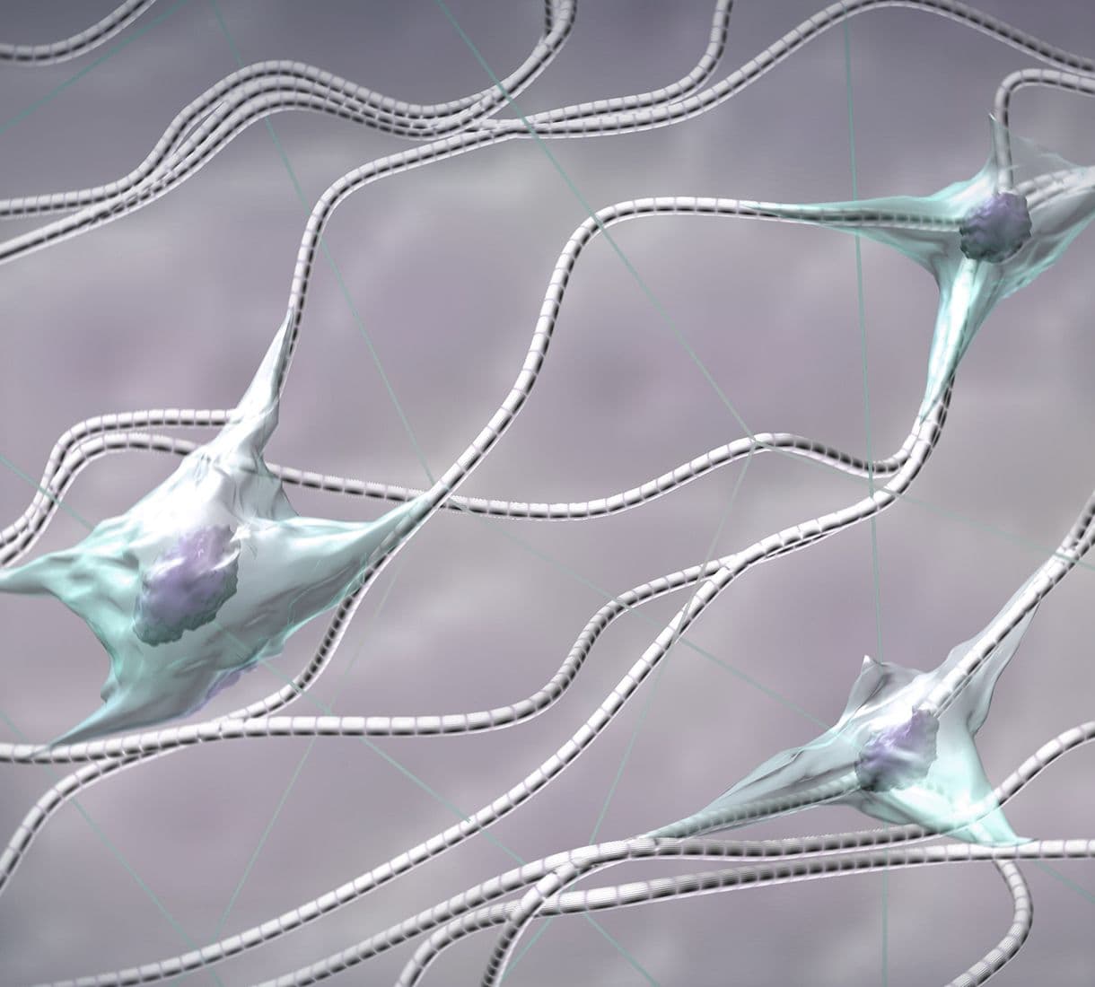
ECM Proteins

The Extracellular Matrix (ECM) is composed of collagen, non-collagenous glycoproteins, and proteoglycans.  These components are secreted from cells to create an ECM meshwork that surrounds cells and tissues. The ECM regulates many aspects of cellular function, including the cells' dynamic behavior, cytoskeletal organization, and intercellular communication.  

Fluorescent_fibronectin_overlay_with_DIC_v3b

MCF10A cells treated with fluorescent rhodamine fibronectin (Cat. # FNR01). Image kindly provided by A. Varadaraj and M. Karthikeyan, Universityof South Carolina, Columbia, SC.

Role in normal cellular functions

Formation of the extracellular matrix (ECM) requires cells to secrete ECM proteins. Assembly is achieved by following a strict hierarchical assembly pattern which begins with the deposition of fibronectin filaments on the cell surface, a process known as fibrillogenesis (1).  Cells continue to remodel the ECM by degradation and reassembly mechanisms, the dynamic nature of the ECM being particularly apparent during development, wound healing, and certain disease states (see below) (2). It is estimated that there are over 300 proteins comprising the mammalian ECM or “core matrisome” and this does not include the large number of ECM-associated proteins (3). Cells interact with the ECM through receptors such as integrins and syndecans, resulting in the transduction of multiple signals to regulate key cellular processes such as differentiation, proliferation, survival, and motility of cells (3).  The ECM has also been shown to bind growth factors such as VEGF, HGF and BMPs which are thought to create growth factor gradients that regulate pattern formation during development (4).  Many of the ECM-regulated cell processes operate via reorganization of the actin and microtubule cytoskeletons (5).

ECM Products


Product 

Cat.#
 Fibronectin
(Red fluorescent, rhodamine) 
FNR01
 

Fibronectin 
(Green fluorescent, HiLyte488TM)

FNR02
 Fibronectin
(Biotinylated)
FNR03
 Laminin 
(Red fluorescent, rhodamine) 
LMN01
 Laminin 
(Green fluorescent, HiLyte488TM
LMN02
 Laminin 
(Biotinylated) 
LMN03

Role in disease

Aberrant regulation or genetic defects of the ECM components often results in a pathogenic state. 

Genetic diseases: Several diseases have been shown to be caused by mutations in ECM genes, including macular degenerative disease (Fibulin 3, ref. 6), osteoarthritis (Asporin, ref. 7) and congenital muscular dystrophy (Laminins, ref. 8). 

Humoral immunity to FBLN-1 has been detected in breast cancer patient sera, indicating that altered expression of a given ECM protein is correlated with cancer (9).  It is also well established that degradation of the ECM, via the actions of matrix metalloproteinases, is a prerequisite to metastatic invasion of cancer cells (10).

Atherosclerosis: This disease has been linked to a buildup of collagen plaques (11).

Biomedical Applications

The field of regenerative medicine and tissue engineering is utilizing ECM components to try to generate a predictable formation of tissues and organs from a given cell type (12,13).

Assays Used to study ECM

There is a preponderance of commercially available ECM reagents and cell attachment/invasion assays. The best known of the ECM reagents is MatrigelTM from BD which is an ECM extract from the Engelbreth-Holm-Swarm mouse tumor. MatrigelTM is composed of laminin (56%), collagen type IV (31%), and enactin (8%), and several growth factors, including EGF (0.7 ng/ml), PDGF (12 ng/ml), IGF-1 (16ng/ml), and TGF-a (2.3 ng/ml) (14).  Similar products include ECMatrixTM (Millipore) and ECM gel (Sigma). Most commercially available invasion assays utilize a Boyden-chamber like system (15).

Cytoskeleton offers a unique line of fluorescently-labeled and biotinylated fibronectins and laminins.  Several applications of these products are listed below.

Application #1:     In vitro invadopodia/podosome invasion assay (Cat.# FNR01, FNR02, LMN01, LMN02)

Cytoskeleton’s fluorescently-labeled fibronectins and laminins can be used in an in vitro invadopodia/podosome invasion assay (16).  This assay allows a high resolution examination of local cell invasion on specific ECM components and can be used to assess the invasive potential of cells and to examine compounds/pathways that affect this stage of invasion.  Originally described by Artym et al. for gelatin, the assay is equally applicable to fluorescent fibronectin or laminin (16).

Application #2:     Signaling pathways involved in fibronectin matrix assembly: fibrillogenesis (Cat.# FNR01, FNR02, FNR03)

Unlike other ECM components that can self-polymerize under physiological conditions, fibronectin matrix assembly is a cell-dependent process. Understanding the mechanisms involved in FN assembly and how these interplay with cellular, fibrotic, and immune responses may reveal targets for the future development of therapies to regulate aberrant tissue-repair processes.  Also, tissue engineering strongly depends on the ability to control the rate and pattern of ECM formation.  Cytoskeleton’s labeled fibronectins can be used to monitor fibrillogenesis;

Fluorescent Fibronectin Substrates (FNR01 & FNR02)

This method involves fluorescent tracing of fibril formation through incorporation of fluorescent fibronectins (17).   

The conversion of soluble fibronectin to insoluble fibrils on the cell surface can be visualized by feeding cell culture with media containing either TRITC-labeled fibronectin (Cat.# FNR01) or HiLyte488-labeled fibronectin (Cat.# FNR02). The level of incorporated fibronectin can be observed and quantitated by fluorescence microscopy (18).

Biotinylated Fibronectin Substrate (FNR03)

This method incorporates biotinylated fibronectin into the cells of interest and quantitates detergent soluble (cell bound) and detergent insoluble (fibrils) fibronectin. This assay has been used successfully to examine the role of Rho proteins in fibrillogenesis (19).

Application #3:     Tissue engineering applications (FNR03 & LMN03)

It has been shown that biotinylated ECM proteins can adopt a more native conformation when bound to a streptavidin surface and that this can lead to enhanced cellular binding to the ECM (20).

References

  1. Sottile J. and Hocking D. 2002. Fibronectin polymerization regulates the composition and stability of extracellular matrix fibrils and cell-matrix adhesions. Mol. Biol. Cell. 13, 3546-3559.
  2. Daley W. et al. 2007. Extracellular matrix dynamics in development and regenerative medicine. J. Cell Sci. 121, 255-264.
  3. Hynes R. and Naba. 2011. Overview of the matrisome-an inventory of extracellular matrix constituents and functions. Cold Spring Harb Perspect Biol. doi:10.1101.
  4. Taipale J. and Keski-Oja J. 1997. Growth factors in the extracellular matrix. FASEB J. 11, 51-59.
  5. Ballestrem C. et al. 2004. Interplay between the actin cytoskeleton, focal adhesions and microtubules. Cell Motility. Ed Anne Ridley, Michelle Peckham and Peter Clark. 75-99.
  6. Klenotic PA. et al. 2004. Tissue inhibitor of metalloproteinases-3 (TIMP-3) is a binding partner of epidermal growth factor-containing fibulin-like extracellular matrix protein 1 (EFEMO1). Implications for macular degeneration. J. Biol. Chem. 279, 30469-30473.
  7. Kizawa H. et al. 2005. An aspartic acid repeat polymorphism in aspirin inhibits chondrogenesis and increases susceptibility to osteoarthritis. Nat. Genet. 37, 138-144.
  8. Hall TE. et al. 2007. The zebrafish candyfloss mutant implicates extracellular matrix adhesion failure in laminin alpha-2-deficient congenital muscular dystrophy. Proc. Natl. Acad. Sci. USA. 104, 7092-7097.
  9. Hu J. et al. 2008.  Matrix metalloproteinase inhibitors as therapy for inflammatory and vascular disease. Nat. Rev. Drug Discov. 6, 480-498.
  10. Pupa S. et al. 2002. New insights into the role of extracellular matrix during tumor onset and progression. J. Cell Physiol. 192, 259-267.
  11. Seyama Y. and Wachi H. 2004. Artherosclerosis and matrix dystrophy. J. Artheroscer. Thromb. 11, 236-245.
  12. Causa F. et al. 2007. A multi-functional scaffold for tissue regeneration: the need to engineer a tissue analog. Biomaterials. 28, 5093-5099.
  13. Barker TH. 2011. The role of ECM proteins and protein fragments in guiding cell behavior in regenerative medicine. Biomaterials. 32, 4211-4214.
  14. Kleinman HK. Et al. 1982. Isolation and characterization of type IV procollagen, laminin and heparin sulfate proteoglycan from EHS sarcoma. Biochemistry. 21, 6188-6193.
  15. Yuan K. et al. 2006. In vitro matrices for studying tumor cell invasion. Cell Motility in Cancer Invasion and Metastasis Ed. A. Wells. 25-54.
  16. Artym V. et al. 2009. ECM degradation assays for analyzing local cell invasion. Meth. Mol. Biol., Extracellular Matrix Protocols. 522, 211-219.
  17. Pankov R. and Momchilova A. 2009. Fluorescent labeling techniques for investigation of fibronectin fibrillogenesis (labeling fibronectin fibrillogenesis). Methods Mol. Biol., Extracellular Matrix Protocols. 522, 261-274. 
  18. Pankov R. et al. 2000.  Integrin dynamics and matrix assembly: tensin-dependent translocation of alpha (5)beta(1) integrins promotes early fibronectin fibrillogenesis. J. Cell Biol. 148, 1075-1090.
  19. Pankov R. and Yamada K. 2004. Non-radioactive quantification of fibronectin matrix assembly. Curr. Protoc. Cell Biol. 10.13.1-10.13.9.
  20. Lehnert M, et al. 2011. Adsorption and conformation behavior of biotinylated fibronectin on streptavidin-modified TiO(X) surfaces studied by SPR and AFM. Langmuir. 27, 7743-7751.

ECM Product Citations

AuthorTitleJournalYearArticle Link
Sadhu, Raj Kumar et al.Experimental and theoretical model for the origin of coiling of cellular protrusions around fibersNature Communications 2023 14:12023ISSN 2041--1723
Gibson, Shayin V. et al.ADAMTS3 restricts cancer invasion in models of early breast cancer progression through enhanced fibronectin degradationMatrix Biology2023ISSN 0945--053X
Dickinson, Richard B. et al.Viscous shaping of the compliant cell nucleusAPL Bioengineering2022ISSN 2473-2877
Van Der Putten, Cas et al.Protein Micropatterning in 2.5D: An Approach to Investigate Cellular Responses in Multi-Cue EnvironmentsACS Applied Materials and Interfaces2021ISSN 1944-8252
Lauko, Domokos I. et al.Baculovirus actin-rearrangement-inducing factor arif-1 induces the formation of dynamic invadosome clustersMolecular Biology of the Cell2021ISSN 1939-4586
Naka, Y. et al.Wholly vascularized millimeter-sized engineered tissues by cell-sized microscaffoldsMaterials Today Bio2020ISSN 2590-0064
Summerbell, Emily R. et al.Epigenetically heterogeneous tumor cells direct collective invasion through filopodia-driven fibronectin micropatterningScience Advances2020ISSN 2375-2548
Sundararaman, Ananthalakshmy et al.RhoJ Regulates α5β1 Integrin Trafficking to Control Fibronectin Remodeling during AngiogenesisCurrent Biology2020ISSN 1879-0445
Lo Vecchio, Simon et al.Collective Dynamics of Focal Adhesions Regulate Direction of Cell MotionCell Systems2020ISSN 2405-4720
Garbett, Damien et al.T-Plastin reinforces membrane protrusions to bridge matrix gaps during cell migrationNature Communications2020ISSN 2041-1723
Roveimiab, Ziba et al.Traction and attraction: Haptotaxis substrates collagen and fibronectin interact with chemotaxis by HGF to regulate myoblast migration in a microfluidic deviceAmerican Journal of Physiology - Cell Physiology2020ISSN 1522-1563
Horvath, Aron N. et al.The Protein Mat(ters) - Revealing the Biologically Relevant Mechanical Contribution of Collagen- And Fibronectin-Coated MicropatternsACS Applied Materials and Interfaces2019ISSN 1944-8252
Tomba, Caterina et al.Laser-Assisted Strain Engineering of Thin Elastomer Films to Form Variable Wavy Substrates for Cell CultureSmall2019ISSN 1613-6829
Rafiq, Nisha Bte Mohd et al.A mechano-signalling network linking microtubules, myosin IIA filaments and integrin-based adhesionsNature Materials2019ISSN 1476-4660
De Vos, Ivo J.H.M. et al.Functional analysis of a hypomorphic allele shows that MMP14 catalytic activity is the prime determinant of the Winchester syndrome phenotypeHuman Molecular Genetics2018ISSN 1460-2083
Hasan, Muhammad M. et al.Invadosome-mediated human extracellular matrix degradation by Entamoeba histolyticaInfection and Immunity2018ISSN 1098-5522
Varadaraj, Archana et al.TGF-β triggers rapid fibrillogenesis via a novel TβRII-dependent fibronectin-trafficking mechanismMolecular Biology of the Cell2017ISSN 1939-4586
Funano, Shun Ichi et al.Vapor-based micro/nano-partitioning of fluoro-functional group immobilization for long-term stable cell patterningRSC Advances2016ISSN 2046-2069
Mana, Giulia et al.PPFIA1 drives active α5β1 integrin recycling and controls fibronectin fibrillogenesis and vascular morphogenesisNature Communications2016ISSN 2041-1723
Blehm, Benjamin H. et al.Deconstructing the role of the ECM microenvironment on drug efficacy targeting MAPK signaling in a pre-clinical platform for cutaneous melanomaBiomaterials2015ISSN 1878-5905
Wood, SheilaDifferentiation of Borrelia Microbes from Collagen Debris and Collagen Fibrils in Blood CulturesJournal of Microbiology & Experimentation2015Article Link
Comelles, Jordi et al.Cells as Active Particles in Asymmetric Potentials: Motility under External GradientsBiophysical Journal2014ISSN 1542-0086
Steele, Amanda N. et al.Tandem zyxin LIM sequences do not enhance force sensitive accumulationBiochemical and Biophysical Research Communications2012ISSN 0006-291X
Nakayama, Masamichi et al.Thermoresponsive poly(N-isopropylacrylamide)-based block copolymer coating for optimizing cell sheet fabricationMacromolecular bioscience2012ISSN 1616--5195
Nagase, Kenichi et al.Thermo-responsive polymer brushes as intelligent biointerfaces: preparation via ATRP and characterizationMacromolecular bioscience2011ISSN 1616--5195
Steward, Robert L. et al.Mechanical stretch and shear flow induced reorganization and recruitment of fibronectin in fibroblastsScientific Reports2011ISSN 2045-2322
Robinson, Elizabeth E. et al.Fibronectin Matrix Assembly Regulates α5β1-mediated Cell CohesionMolecular Biology of the Cell2004ISSN 1059-1524
Brock, Amy et al.Geometric Determinants of Directional Cell Motility Revealed Using Microcontact Printing†Langmuir2003ISSN 0743-7463
AuthorTitleJournalYearArticle Link
Xia, Qiaoxi et al.A protein complex of LCN2, LOXL2 and MMP9 facilitates tumour metastasis in oesophageal cancerMolecular Oncology2023ISSN 1878--0261
Baschieri, Francesco et al.Fibroblasts generate topographical cues that steer cancer cell migrationScience Advances2023ISSN 2375-2548
Han, Zuoning et al.Integrin aVβ1 regulates procollagen i production through a non-canonical transforming growth factor β signaling pathway in human hepatic stellate cellsBiochemical Journal2021ISSN 1470-8728
Pal, Kaushik et al.Ubiquitous membrane-bound DNase activity in podosomes and invadopodiaJournal of Cell Biology2021ISSN 1540-8140
Li, Wenhong et al.Differential cellular responses to adhesive interactions with galectin-8- And fibronectin-coated substratesJournal of Cell Science2021ISSN 1477-9137
Rong, Zhouyi et al.Activation of FAK/Rac1/Cdc42-GTPase signaling ameliorates impaired microglial migration response to Aβ42 in triggering receptor expressed on myeloid cells 2 loss-of-function murine modelsFASEB Journal2020ISSN 1530-6860
Huang, Yuxing et al.Arp2/3-branched actin maintains an active pool of GTP-RhoA and controls RhoA abundanceCells2019ISSN 2073-4409
Stanton, Alice E. et al.Biochemical Ligand Density Regulates Yes-Associated Protein Translocation in Stem Cells through Cytoskeletal Tension and IntegrinsACS Applied Materials and Interfaces2019ISSN 1944-8252
Foxall, Elizabeth et al.PAK4 Kinase Activity Plays a Crucial Role in the Podosome Ring of Myeloid CellsCell Reports2019ISSN 2211-1247
Sala, Laura et al.Abrogation of myofibroblast activities in metastasis and fibrosis by methyltransferase inhibitionInternational Journal of Cancer2019ISSN 1097-0215
Rafiq, Nisha Bte Mohd et al.Forces and constraints controlling podosome assembly and disassemblyPhilosophical Transactions of the Royal Society B: Biological Sciences2019ISSN 1471-2970
Sun, Xiaoyu et al.Replication of biocompatible, nanotopographic surfacesScientific Reports2018ISSN 2045-2322
Werley, Christopher A. et al.Geometry-dependent functional changes in iPSC-derived cardiomyocytes probed by functional imaging and RNA sequencingPLoS ONE2017ISSN 1932-6203
Kim, Jiyun et al.Three-dimensional patterning of the ECM microenvironment using magnetic nanoparticle self assemblyCurrent Protocols in Cell Biology2016ISSN 1934-2616
Kim, Jiyun et al.Independent Control of Topography for 3D Patterning of the ECM MicroenvironmentAdvanced Materials2016ISSN 1521-4095
Torr, Elizabeth E. et al.Myofibroblasts exhibit enhanced fibronectin assembly that is intrinsic to their contractile phenotypeJournal of Biological Chemistry2015ISSN 1083-351X
Stanisavljevic, Jelena et al.Snail1-expressing fibroblasts in the tumor microenvironment display mechanical properties that support metastasisCancer Research2015ISSN 1538-7445
Lively, Starlee et al.The microglial activation state regulates migration and roles of matrix-dissolving enzymes for invasionJournal of Neuroinflammation2013ISSN 1742-2094
Jacob, Abitha et al.Rab40b regulates trafficking of MMP2 and MMP9 during invadopodia formation and invasion of breast cancer cellsJournal of cell science2013ISSN 1477--9137
AuthorTitleJournalYearArticle Link
Phillips, Andrew T. et al.USP10 promotes fibronectin recycling, secretion, and organizationInvestigative Ophthalmology and Visual Science2021ISSN 1552-5783
Varadaraj, Archana et al.Deoxycholate Fractionation of Fibronectin (FN) and Biotinylation Assay to Measure Recycled FN Fibrils in Epithelial CellsBio-Protocol2018ISSN 2331--8325
Varadaraj, Archana et al.TGF-β triggers rapid fibrillogenesis via a novel TβRII-dependent fibronectin-trafficking mechanismMolecular Biology of the Cell2017ISSN 1939-4586
Wang, Qiang et al.The human blood parasite Schistosoma mansoni expresses extracellular tegumental calpains that cleave the blood clotting protein fibronectinScientific Reports2017ISSN 2045-2322
Rodriguez, Natalia M. et al.Micropatterned multicolor dynamically adhesive substrates to control cell adhesion and multicellular organizationLangmuir2014ISSN 0743-7463
AuthorTitleJournalYearArticle Link
Frenzel, Jan et al.Laminin Adsorption and Adhesion of Neurons and Glial Cells on Carbon Implanted Titania Nanotube Scaffolds for Neural Implant ApplicationsNanomaterials 2022, Vol. 12, Page 38582022ISSN 2079--4991
Coelho-Sampaio, Tatiana et al.Type IV collagen conforms to the organization of polylaminin adsorbed on planar substrataActa Biomaterialia2020ISSN 1878-7568
Zeng, Jinfeng et al.In Situ Cross-Linking of Artificial Basement Membranes in 3D Tissues and Their Size-Dependent Molecular PermeabilityBiomacromolecules2020ISSN 1526-4602
Fiore, Vincent F. et al.Mechanics of a multilayer epithelium instruct tumour architecture and functionNature2020ISSN 1476-4687
Zeng, Jinfeng et al.Fabrication of Artificial Nanobasement Membranes for Cell Compartmentalization in 3D TissuesSmall2020ISSN 1613-6829
Maechler, Florian A. et al.Curvature-dependent constraints drive remodeling of epitheliaJournal of Cell Science2019ISSN 1477-9137
Melero, Cristina et al.Light-induced molecular adsorption of proteins using the primo system for micro-patterning to study cell responses to extracellular matrix proteinsJournal of Visualized Experiments2019ISSN 1940-087X
Alessandri, Kevin et al.A 3D printed microfluidic device for production of functionalized hydrogel microcapsules for culture and differentiation of human Neuronal Stem Cells (hNSC)Lab on a Chip2016ISSN 1473--0189
Lantoine, Joséphine et al.Matrix stiffness modulates formation and activity of neuronal networks of controlled architecturesBiomaterials2016ISSN 1878-5905
Kim, Jiyun et al.Three-dimensional patterning of the ECM microenvironment using magnetic nanoparticle self assemblyCurrent Protocols in Cell Biology2016ISSN 1934-2616
Kim, Jiyun et al.Independent Control of Topography for 3D Patterning of the ECM MicroenvironmentAdvanced Materials2016ISSN 1521-4095
Hinüber, C. et al.Hierarchically structured nerve guidance channels based on poly-3-hydroxybutyrate enhance oriented axonal outgrowthActa Biomaterialia2014ISSN 1742--7061
AuthorTitleJournalYearArticle Link
Mattiassi, Sabrina et al.The Combined Effects of Topography and Stiffness on Neuronal Differentiation and Maturation Using a Hydrogel PlatformCells2023ISSN 2073-4409
Coelho-Sampaio, Tatiana et al.Type IV collagen conforms to the organization of polylaminin adsorbed on planar substrataActa Biomaterialia2020ISSN 1878-7568

Question 1:  Can I coat cells with fluorescent ECM proteins?

Answer 1: Yes, cells can be coated with fluorescently-labeled ECM proteins such as fibronectin.    Example protocols are found in Pallis et al., 1997 (Peripheral Blood Lymphocyte Binding to a Soluble FITC-Fibronectin Conjugate. Cytometry. 28, 157-164) and Huveneers et al., 2008 (Binding of soluble fibronectin to integrin a5b1 – link to focal adhesion redistribution and contractile shape. J. Cell Sci. 121, 2452-2462).  Briefly, cells are incubated with a low concentration of soluble fluorescently-labeled fibronectin in an appropriate buffer.  Low concentrations of fibronectin are recommended to minimize non-specific binding.  The cells/fibronectin mixture is incubated in the dark for 30 min at room temperature.  Others have done the incubation at 4°C for 1 hour.  The cells are then rinsed in an appropriate buffer, resuspended in the same buffer, and then the coating/binding of ECM proteins can be quantified by flow cytometry or the coated cells can be used experimentally.

Select from the categories below: