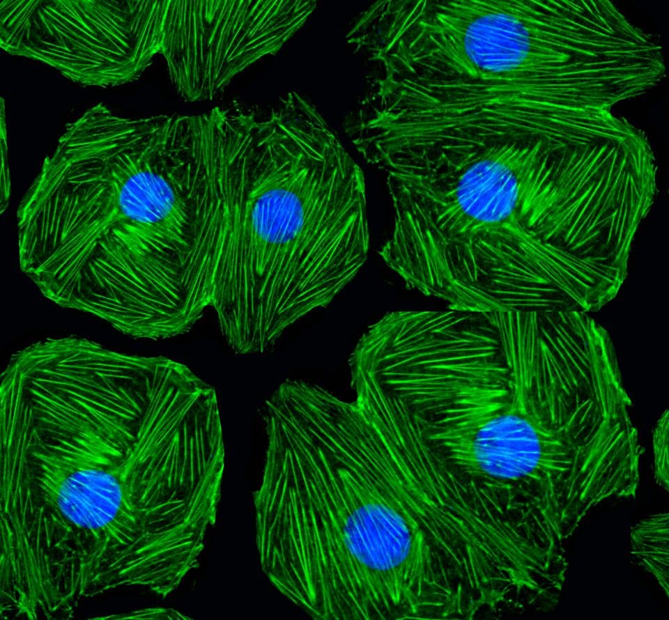
The Acti-stain™ line of fluorescent phalloidins has been developed with an emphasis on creating exceptionally bright and stable probes at an economical price. Side-by-side comparisons with leading competing products demonstrate that you will enjoy savings while not sacrificing the quality of the stain when switching to an Acti-stain™ probe. Additionally, these probes are compatible with popular filter sets such as FITC, TRITC and Cy 5. The combination of in-house manufacturing, stringent quality control and convenient packaging provides a great value. Give them a try and see for yourself.
Product Uses Include
Actin staining is very useful in determining the structure and function of the cytoskeleton in living and fixed cells. The actin cytoskeleton is a very dynamic and labile structure in the living cell, but it can be fixed with paraformaldehyde prior to probing or staining for actin structures.
Click here for Acti-stain specifications
Click here for General F-actin staining information and protocols
Cytoskeleton's Acti-stain products have been cited many times since their introduction. A select few are described here, for more citations on individual products please use the "Citations" tab on each individual product page.
Ezratty, E. J., Partridge, M. A. and Gundersen, G. G. (2005). Microtubule-induced focal adhesion disassembly is mediated by dynamin and focal adhesion kinase. Nat. Cell Biol. 7, 581-590.
Gomes, E. R., Jani, S. and Gundersen, G. G. (2005). Nuclear movement regulated by Cdc42, MRCK, myosin, and actin flow establishes MTOC polarization in migrating cells. Cell 121, 451-463.
Roitbak et al., 2011. The role of microRNAs in neural stem cell supported endothelial morphogenesis. Vascular Cell. v 3, p 25.
Yuan et al., 2012. Subsecond absolute quantitation of amine metabolites using isobaric tags for discovery of pathway activation in mammalian cells. Anal. Chem. v 84, pp 2892–2899.
Andersen et al., 2012. Escherichia coli Uropathogenesis In Vitro: Invasion, Cellular Escape, and Secondary Infection Analyzed in a Human Bladder Cell Infection Model. Infect. Immun. v 80, pp 1858-1867.
Cuajungco et al., 2012. Abnormal accumulation of human transmembrane (TMEM)-176A and 176B proteins is associated with cancer pathology. Acta Histochemica. v 114, pp 705-712.
Ahrens et al., 2012. F-Actin Is an Evolutionarily Conserved Damage-Associated Molecular Pattern Recognized by DNGR-1, a Receptor for Dead Cells. Immunity. v 36, pp 635-645.
Question 1: Which is the most stable/brightest Acti-stain conjugate?
Answer 1: The brightest and most stable of the Acti-stains is Acti-stain 488 (green fluorescence; Cat. # PHDG1). Please see the table below for additional information on all of our Acti-stains.
* = measured in the absence of antifade.
Question 2: What fixation conditions are recommended when staining with phalloidin conjugates?
Answer 2: Fluorescent phalloidins only bind to the native quaternary structure of F-actin which provides a low background. The correct fixation condition for phalloidin binding is 3.7% (v/v) paraformaldehyde in PBS for 10 min because it retains the quaternary protein structure which is necessary for high affinity binding of phalloidin. Methanol fixation destroys the native conformation and hence is not suitable for F-actin staining with phalloidin.
For more information about staining procedures and specifications of the Acti-stain series, please click here.