Tubulin Assays

Since 1993 Cytoskeleton has provided purified tubulin proteins to the scientific community. We continually strive to provide the purest, most biological active and relevant tubulin proteins, kits and reagents for today's researchers. The kits are designed to enhance the productivity ot the time you have available for research, usually saving months of development time. Kits range from the basic tubulin protein and buffers version, BK015, to the Microtubule Binding Assay, BK029, and the polymerization assays described below. We also provide a kit for producing fluorescent microtubules, BK007R, and one for measuring the ratio of microtubules to subunit tubulin in cell extracts BK038. Detailed information can be found by clicking on the links below.

The Microtubule Binding Spin-down Assay, BK029, uses pre-formed microtubules as a substrate to bind the protein or compound of interest, the bound and non-bound fractions are separated by centrifugation to produce a pellet (bound) and supernatant (non-bound) fractions.

MT_schematic_2

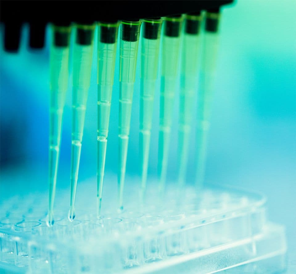
There are two types of tubulin polymerization assay, these are the absorbance and fluorescence formats. In most cases the fluorescence format is recommended because the assay is highly sensitive and economical. The optical density based tubulin polymerization assay (Cat. # BK004P) is highly referenced in scientific literature. The BK006P tubulin polymerization assay uses highly purified tubulin, which is a good secondary screen for determining IC50s and tubulin specificity. The fluorescence tubulin polymerization assay (Cat. # BK011P) is very robust and is well cited in the literature (click on the Citations Tab to see the references to this product).


For more selective anti-cancer drug development a very exciting secondary screen can be achieved using Cancer Cell Line Tubulin. Currently, HeLa and MCF-7 cell line tubulins can be used in the fluorescence polymerization assay (Cat. # H001 and H005, respectively). It has been shown that anti-cancer drugs have different affinities for these tubulins compared to bovine brain (neuronal) tubulin. This indicates that a drug development program aimed at increasing the affinity for cancer cell line tubulin over neuronal tubulin may lead to greater targeting efficiency toward cancer cells.


We also provide HTS assays for other types of tubulins, such as fungal tubulins, plant tubulins, or bacterial tubulin analogs. If you are interested in assaying a specific tubulin not listed on our website, please contact our technical support to discuss how we can provide you with the specific tubulin type you want.

Many publications cite the use of Cytoskeleton's kits in the Materials and Methods section of papers. Usually the citation is associated with a particular result in the form of a graph or image that helps the authors present their findings. This indicates the utility of the Kits to produce publication quality data in a short timeframe thus helping improve the productivity of your efforts. Example citations for tubulin assay kits are shown below.  More citations are available on individual product pages.


Tubulin polymerization HTS assay using >99% pure tubulin, OD based - Porcine  (Cat. # BK006P)

O'Boyle NM, Carr M, Greene LM, Bergin O, Nathwani SM, McCabe T, Lloyd DG, Zisterer DM, Meegan MJ. (2010). Synthesis and Evaluation of Azetidinone Analogues of Combretastatin A-4 as Tubulin Targeting Agents.. J Med Chem.
Jacob Kushkuley, Walter K. H. Chan, Sangmook Lee, Joel Eyer, Jean-Francois Leterrier, Franck Letournel and Thomas B. Shea (2009). Neurofilament cross-bridging competes with kinesin-dependent association of neurofilaments with microtubules. J Cell Science 122 ,3579-86.
Chen, Z., Merta, P. J., Lin, N. H., Tahir, S. K., Kovar, P., Sham, H. L. and Zhang, H. (2005). A-432411, a novel indolinone compound that disrupts spindle pole formation and inhibits human cancer cell growth. Mol. Cancer Ther. 4, 562-568.
Huang, Y. T., Huang, D. M., Guh, J. H., Chen, I. L., Tzeng, C. C. and Teng, C. M. (2005). CIL-102 interacts with microtubule polymerization and causes mitotic arrest following apoptosis in the human prostate cancer PC-3 cell line. J. Biol. Chem. 280, 2771-2779.
Rouzier, R., Rajan, R., Wagner, P., Hess, K. R., Gold, D. L., Stec, J., Ayers, M., Ross, J. S., Zhang, P., Buchholz, T. A. et al. (2005). Microtubule-associated protein tau: A marker of paclitaxel sensitivity in breast cancer. Proc. Natl. Acad. Sci. U. S. A. 102, 8315-8320.
Jiang, J. D., Denner, L., Ling, Y. H., Li, J. N., Davis, A., Wang, Y., Li, Y., Roboz, J., Wang, L. G., Perez-Soler, R. et al. (2002). Double blockade of cell cycle at G1-S transition and M phase by 3-iodoacetamido benzoyl ethyl ester, a new type of tubulin ligand. Cancer Res. 62, 6080-6088.
Mooberry, S. L., Tien, G., Hernandez, A. H., Plubrukarn, A. and Davidson, B. S. (1999). Laulimalide and isolaulimalide, new paclitaxel-like microtubule-stabilizing agents. Cancer Res. 59, 653-660.

Fluorescent Microtubules Biochem Kit  (Cat. # BK007R)

Tong, T., Ji, J., Jin, S., Li, X., Fan, W., Song, Y., Wang, M., Liu, Z., Wu, M. and Zhan, Q. (2005). Gadd45a expression induces Bim dissociation from the cytoskeleton and translocation to mitochondria. Mol. Cell Biol. 25, 4488-4500.

Microtubules/Tubulin Biochem Kit  (Cat. # BK015)

van Horck, F. P., Ahmadian, M. R., Haeusler, L. C., Moolenaar, W. H. and Kranenburg, O. (2001). Characterization of p190RhoGEF, a RhoA-specific guanine nucleotide exchange factor that interacts with microtubules. J. Biol. Chem. 276, 4948-4956.

Microtubule Binding Protein Spin-Down Assay Biochem Kit  (Cat. # BK029)

Cho, H. P., Liu, Y., Gomez, M., Dunlap, J., Tyers, M. and Wang, Y. (2005). The dual-specificity phosphatase CDC14B bundles and stabilizes microtubules. Mol. Cell. Biol. 25, 4541-4551.
Monzo, P., Gauthier, N. C., Keslair, F., Loubat, A., Field, C. M., Le Marchand-Brustel, Y. and Cormont, M. (2005). Clues to CD2-associated Protein Involvement in Cytokinesis. Mol. Biol. Cell 16, 2891-2902.
Lansbergen, G., Komarova, Y., Modesti, M., Wyman, C., Hoogenraad, C. C., Goodson, H. V., Lemaitre, R. P., Drechsel, D. N., van Munster, E., Gadella, T. W., Jr. et al. (2004). Conformational changes in CLIP-170 regulate its binding to microtubules and dynactin localization. J. Cell Biol. 166, 1003-1014.
Pennetta, G., Hiesinger, P., Fabian-Fine, R., Meinertzhagen, I. and Bellen, H. (2002). Drosophila VAP-33A directs bouton formation at neuromuscular junctions in a dosage-dependent manner. Neuron 35, 291-306.
Ziegelbauer, J., Shan, B., Yager, D., Larabell, C., Hoffmann, B. and Tjian, R. (2001). Transcription factor MIZ-1 is regulated via microtubule association. Mol. Cell 8, 339-349.

Microtubule/Tubulin In Vivo Assay Biochem Kit  (Cat. # BK038)

Davis, F. J., Pillai, J. B., Gupta, M. and Gupta, M. P. (2005). Concurrent opposite effects of trichostatin A, an inhibitor of histone deacetylases, on expression of ƒ¿-MHC and cardiac tubulins: implication for gain in cardiac muscle contractility. Am. J. Physiol. 288, H1477-1490.

Question 1: What is the best way to test whether my protein interacts with tubulin?

Answer 1:  To examine if your protein of interest interacts with tubulin, we recommend using one of our tubulin polymerization assays (Cat. # BK004P, BK006P or BK011P) for a sensitive measure of how the protein affects one or more of the three phases of polymerization (nucleation, growth and steady state).  If microtubule is suspected, we recommend confirming that there are direct interactions between microtubules and your protein of interest with the Microtubule Binding Protein Spin-down Assay Biochem Kit (Cat. # BK029). If tubulin heterodimer subunit binding is suspected (i.e. polymerization is inhibited) then one can coat streptavidin 96-well plates with biotinylated tubulin (Cat. # T333P) to create a tubulin subunit capture surface.  Then the compound or protein can be incubated in the well and binding can be visualized by an HRP detection system using an antibody (or a tag can be conjugated to the test protein) against the compound or protein of interest.  Keep in mind that buffer composition is also important as it will need to be low salt (approx 50 mM) and contain MgCl2 (or other divalent cation) and GTP to maintain subunit integrity.  For an example of a compatible buffer, see our recipe for general actin buffer (Cat. # BST01-001).  Alternatively, the protein of interest could be tagged with GST (avoid reducing agents) and conjugated to the plate and then incubated with biotinylated tubulin and visualized with HRP-conjugated streptavidin.


Question 2: What equipment do I need for the microtubule binding assay?

Answer 2:  Equipment required for the assay are an ultracentrifuge capable of centrifuging 50-200 μl volumes at 100,000 x g at 4°C and 24°C is needed.  Examples of such centrifuges are the Beckman airfuge with ultraclear tubes (Beckman, cat. # 344718), the SW50 ultracentrifuge rotor with adapters for ultraclear tubes (Beckman, Cat. # 344718) and a Beckman tabletop ultracentrifuge with TLA-100 rotor. 

Reagents and components not supplied with the kit include:  Laemmli reducing-sample buffer (1x stock, 62.5 mM Tris-HCl pH 6.8, 2% SDS, 10% glycerol, 0.001% bromophenol blue and 5% v/v β-mercaptoethanol) and (5x stock, 300 mM Tris-HCl pH 6.8, 10% SDS, 50% glycerol, 0.005% bromophenol blue and 25% v/v β-mercaptoethanol), polyacrylamide gels (12% or 4-20% gradient gels), SDS-PAGE apparatus and buffers, reagents for the detection of the “test” protein (Coomassie, silver stain or antibody), 50% (v/v) glycerol in Milli-Q water for Cushion Buffer resuspension and TCA solution (50% w/v) in Milli-Q water for protein precipitation if necessary.

The kit contents include: tubulin protein (Cat. # T240), MAP fraction (Cat. # MAPF), BSA protein, general tubulin buffer (Cat. # BST01), cushion buffer (Cat. # BST05-001), salt extraction buffer, GTP stock (Cat. # BST06), taxol stock (Cat. # TXD01) and anhydrous DMSO.


Question 3: What is a good negative control for the microtubule binding reaction?

Answer 3:  Actin filaments are a good negative control because they are also negatively charged and pellet like the microtubules under high speed centrifugation.  Confirming that your protein of interest does not bind to actin filaments, but does bind to microtubules is additional confirmation that the test protein is a microtubule-associated protein (MAP).


For more information, click on the Documents tab above, or for Technical Support email tservice@cytoskeleton.com.

  1. Tubulin polymerization HTS assay using >97% pure tubulin, OD based - Porcine (BK004P) BK004P
    Tubulin polymerization HTS assay using >97% pure tubulin, OD based - Porcine (BK004P)
    Learn More
  2. Tubulin polymerization assay using >99% pure tubulin, OD based - Porcine (BK006P) BK006P
    Tubulin polymerization HTS assay using >99% pure tubulin, OD based - Porcine (BK006P) Learn More
  3. Tubulin polymerization assay using >99% pure tubulin, fluorescence based (BK011P) BK011
    Tubulin polymerization HTS assay using >99% pure tubulin, fluorescence based (BK011P)
    Learn More
  4. Microtubule Binding Protein Spin-Down Assay Biochem Kit BK029
    Microtubule Binding Protein Spin-Down Assay Biochem Kit
    Learn More
  5. Microtubule/Tubulin In Vivo Assay Biochem Kit BK038
    Microtubule/Tubulin In Vivo Assay Biochem Kit
    Learn More