Small G-proteins

Cytoskeleton, Inc. has the widest line of small G-protein related products on the market. Cytoskeleton sells purified proteins, a large number of Biochem Kits™ to assay the biology and biochemistry of small G-proteins as well as antibodies to G-proteins and related proteins. See each individual product page for examples of uses.  Bulk quantities of all these proteins are available for substantial discounts.

For more information about Small G-proteins please click the About tab above.

Overview

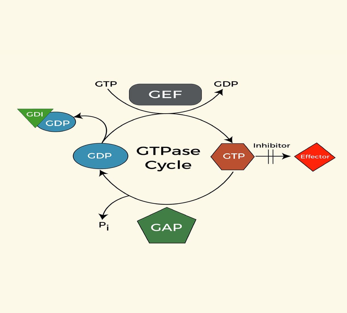
The Ras superfamily of small GTPases consist of more than 150 members, which based on their sequence homology, are divided into several subfamilies such as Rho, Ras, Ran, Rab, Arf and Rad/Rem/Gem/Kir. This group of small GTPases serve as binary switches cycling between GDP-bound inactive and GTP-bound active state (1,2).  The regulatory proteins for this switch include guanine nucleotide exchange factors (GEFs), GTPase activating proteins (GAPs) and guanine nucleotide dissociation inhibitors (GDIs) (1,2).

The Rho subfamily

The Rho family of small GTPases consists of at least 20 members, the most extensively characterized of which are the Rac1, RhoA and Cdc42 proteins (3).  In common with all other small GTPases, the Rho proteins act as molecular switches that transmit cellular signals through an array of effector proteins. This family is involved in a wide range of cellular responses, including cytoskeletal reorganization (4-5), regulation of transcription (6), cell migration (7), cellular transformation and metastasis (8).

The Ras subfamily

There are approximately 10 members of this subfamily split between Ras, Ral and Rap proteins. Ras proteins function in the Raf/ERK signaling pathway which controls proliferation and differentiation. Ral proteins function in the early steps of endocytosis, whereas Rap’s function as antagonist of Ras and they are located to the late endosomal and early lysosomal compartments.

The Ran subfamily

The Ran proteins regulate nuclear transport of macromolecules, and in the cell cycle checkpoint from DNA replication to entry into mitosis.  

The Rab subfamily

One of the largest subfamilies of small G-proteins, the Rab proteins regulate the flow of vesicles from the ER to the Golgi and onto the plasma membrane. They are ubiquitously expressed and are highly abundant.  

The Rad/Rem/Gem/Kir subfamily

Members of this subfamily (RGK subfamily) have wide ranging functions such as controlling cardiac hypertrophy, inhibiting insulin-stimulated glucose uptake in myocyte and adipocyte cell lines, as well as inhibiting voltage dependent calcium channels by binding to the b-subunit (9).

The Arf subfamily

The Arf family consists of Arf proteins and Arf-like proteins (Arl’s) (10). As a general rule the Arf proteins help regulate vesicle trafficking and membrane fusion in functions such as nuclear membrane fusion, negative regulation of Golgi vesicle transport, and plasma membrane invagination. This family is the most divergent of the small G-proteins being equally related to heterotrimeric G-protein family. 


Small G-protein measurement

There are several methods used to measure Small G-proteins, these include a) measurement of the active "GTP-bound" form in extracts and in situ, b) measuring total Small G-protein levels, c) Exchange activity from GTP to GDP and vice versa, and d) the endogenous GTPase rate.  Cytoskeleton provides kits for all these assays (e.g. BK124, BK100, BK100 and BK105).

  1. Measuring the active GTP-bound form of Rho: Traditionally, this assay has been performed using a pull-down method, wherein the Rho-GTP- binding domain (RBD) of a Rho effector is coupled to agarose beads, allowing affinity based detection of the active Rho in biological samples (11). This method suffers from several drawbacks such as being time consuming, requiring large amount of total cellular protein, being limited in the number of samples that can be handled simultaneously and yielding only semi-quantitative results. Cytoskeleton brought this concept to a new level of accuracy and sensitivity by introducing the GLISA technology which uses 96-well format and small amounts of cells/protein and provides highly accurate results. (more details are described here).
  2. Measuring total Small G-protein levels:Once assumed to be an insignificant bystander in cell signalling the total level of Small G-proteins has become a real factor in normal and diseased cell function (12, Burridge 2008). Using either a western blot approach (e.g. with Cat. # ARH03), or a sandwich ELISA (e.g., Cat.# BK150) it is possible to measure total Rho levels in a cell or tissue extract. 
  3. Measuring GEF exchange activity:  Using GTP fluorescent conjugates, which show differential fluorescence upon binding Small G-proteins, one can determine the rate of exchange of GTP, for more information see Cat. #BK100. This assay can be performed on purified recombinant proteins or by immunoprecipitating Rho protein from a cell extract (see the paper referenced in BK100 FAQ Question 1: BK100). 
  4. Measuring endogenous GTPase activity: Although it is difficult to measure GTPase activity from cell extracts because of competing non-specific phosphatase activity, it is relatively straightforward to measure it from a purified recombinant protein with a phosphate assay such as Cat. # BK105 or BK054 offered by Cytoskeleton.

    Guanine exchange factors

    GEFs catalyze the exchange of GDP for GTP to generate the active state of small GTPases in response to extracellular signals. In order to facilitate the exchange, the GEFs must bind to the GDP bound GTPases, destabilize the GDP-GTPase complex, and then stabilize a nucleotide free reaction intermediate. Because of the high intracellular ratio of GTP to GDP, the released GDP is replaced with GTP, leading to release of GEFs from the complex and activation of the GTPase (1,2). Many GEF proteins have been identified as oncogenes and are involved in human disease such as cancer. Interestingly, the expression of GEF protein is tissue or cell type specific, providing a therapeutic potential for cancer treatment (2, 13).

    Recently developed fluorescence analogs of guanine nucleotides have greatly improved the technical ability to define the real time exchange reaction of GEFs, including kinetic and thermodynamic properties, eliminating the need for traditional radioactive labeling method (14, 15). This fluorescence based assay takes advantage of the spectroscopic difference between bound and unbound fluorescent analogs to guanine nucleotides and thus is able to monitor the states of small GTPases (14, 15). One of most widely used fluorescent analogs is mant (N-methylanthraniloyl) fluorophores. Excitation of mant flurophore at 360nm (+/-10nm) gives a fluorescence emission at 440nm (+/-20nm). Once bound to GTPases, the fluorophore emission intensity increases dramatically approximately 2 fold. Therefore, the enhancement of fluorescent intensity in the presence of small GTPases and GEFs will reflect the respective GEF activities of known or unknown proteins.

    Cytoskeleton Inc. has developed a Mant fluorophore based GEF assay suitable for both 96-well and 384-well format. This assay can be applied to multiple research purposes such as characterizing the GEFs and identifying GEF inhibitors in a high throughput screen format. This kit contains human Cdc42, Rac1 and RhoA proteins and the GEF domain of Dbs as a positive control GEF for Cdc42 (Fig. 1) and RhoA.  Dbs shows extremely low GEF activity for Rac1 (see Fig.2 and ref. 4). Interestingly, it was reported that human Dbs can activate Rac1 in a FRET based assay (16).

    BK100

    Legend: The small GTPases Cdc42 (Cat# CD01) and the human Dbs protein (Cat# GE01) were expressed as His-tagged proteins in E. coli and purified on a nickel affinity column.  The reactions were conducted in a 96 well black flat bottom half area plate (Corning Cat# 3686) format (150 µl reaction volumes).  Each reaction contains 1 µM GTPases, 50 µg/ml bovine serum albumin (BSA), 20 mM Tris pH 7.5, 50 mM NaCl, 10 mM MgCl2, and 0.8 µM mant-GTP with or without the presence of 0.5 µM human Dbs(DH/PH) protein. Reactions were measured in a Tecan Spectrofluor plus fluorimeter(lex=360nm, lem=460nm). Readings were taken at 20°C every 30 seconds for a total reaction time of 30 minutes.


    GTPase Activating Proteins

    The balance of the GTP to GDP bound state underlies the switch mechanism as they turn from an activated (GTP form) to inactive state (GDP form). The balance of GTP to GDP bound states is controlled by catalytic proteins that either increase the rate of exchange of GDP for GTP (GEFs), increase the GTPase activity (GAPs), or prevent the exchange of GDP (GDIs). Recently it has been shown that the GAP family of proteins is large (70 members) and potentially important for changing a cell from a normal to disease status (see review in ref. 17).

    GAP activity can be reduced by deletion or mutation in cells; in this case the small GTPase targeted by the GAP has an extended time in the active GTP state, in essence creating a permanently active state. For example the ability of Rho GTPase to control axonal guidance is exemplified in the finding that mutations of a Rho-GAP can lead to mental retardation (18,19). In cancer, mutated Ras-GAPs (NF1 gene) have been shown to cause neurofibromatosis (20,21), and mutated Rheb-GAP has been shown to cause tuberous sclerosis complex (21).

    Although some GAPs are known to have GAP activity, the majority of assumed GAP proteins have only been implicated by homology to contain GAP activity. Cytoskeleton Inc. is facilitating the exploration of this field by introducing the GAP Assay Kit (Cat. # BK105). Several small GTPase proteins (Ras, RhoA, CDC42 and Rac1) are included such that the researcher can screen the small G-proteins for GAP like activity which is usually small G-protein specific i.e. RhoGAP or Ras GAP. It is likely that new domain information will identify other small G-protein GAPs which have not been apparent. In addition the new assay kits are easily adapted for High Throughput Screen format which allows development of ligands for pharmaceutical studies. Please inquire for bulk reagent quotes if you are considering a screening application (tservice@cytoskeleton.com).

     The reagents in this kit have been optimized to enable high activity from your GAP protein such that you can detect the enhanced GTPase of a small G-protein through a simple absorbance based detection method. The small G-protein is incubated in the presence of GAP protein (Rho GAP is the control protein included in this kit), GTP and the optimized buffer. The overall GTPase activity of small G-proteins is composed of two components which limit the activity; these are a) endogenous GTPase activity and b) GDP dissociation rate. The endogenous activity can be enhanced by the addition of a suitable GAP protein, whereas the dissociation rate can be enhanced by buffer optimization.       


    Figure 2.  p50 RhoGAP activity measured by GTP hydrolysis by RhoA and Rac1 protein

    BK105

    Method: The small GTPases RhoA (Cat. # RH01), Rac1 (Cat. # RC01) and the human p50 RhoGAP protein (Cat. # GAS01) were expressed and purified from E. coli and are available as separate items from Cytoskeleton Inc.  The reactions were conducted in a half area clear 96 well plate (Corning Cat. # 3696) format (40 µl reaction volumes).  Each reaction contains +/- 5 µg RhoA or +/- Rac1, +/- 8 µg p50 RhoGAP domain and 200 µM GTP in Reaction Buffer. Sample 1 contained Rac1 only, sample 2 contained Rac1 and p50 RhoGAP, sample 3 contained RhoA only, sample 4 contained RhoA and p50 RhoGAP, and sample 5 contained p50 RhoGAP alone.  Reactions were incubated at 37ºC for 20 min followed by the addition 120 µl of CytoPhos reagent for 10 min to determine the phosphate generated by the hydrolysis of GTP.


    References

    1. Shielge, J. M., et al. Trend Cell Biol., 2000, 10, 147-54.
    2. Whitehead, I. P., et al. Biochim. Biophys. Acta,1997, 1332, F1-23.
    3. Jaffe, AB. & Hall, A. Rho GTPases: Biochemistry and Biology.  Ann. Rev. Cell Dev. Biol. 21: 247-269 (2005)
    4. Ridley, AJ. & Hall, A. The small GTP-binding protein rho regulates the assembly of focal adhesions and actin stress fibers in response to growth factors. Cell 70: 389-399 (1992)
    5. Ridley, AJ. et al. The small GTP-binding protein Rac regulates growth factor-induced membrane ruffling. Cell 70: 401-410 (1992)
    6. Coso, OA., et al.  The small GTP-binding proteins Rac and Cdc42 regulate the activity of the JNK/SAPK signaling pathway.  Cell 81: 1137-1146 (1995)
    7. Small, JV., et al. The lamellipodium: where motility begins.  Trends Cell Biol.12: 112-120 (2002)
    8. Jaffe, A, & Hall, A,  Rho GTPases in transformation and metastasis, Adv. Cancer Res. 84: 57-80 (2002)
    9. Chang L. et al. 2007. Rad GTPase Deficiency Leads to Cardiac Hypertrophy. Molecular Cardiology, 116: 2976-2983.
    10. Clark et al. 1993. Selective amplification of novel members of the ADF-ribosylation factor (Arf) family: cloning of new human and Drosophila ARL genes. PNAS, 90, 8952-6.
    11. Herrmann, C., Martin, G.A. and Wittinghofer, J. Biol. Chem. 270: 2901 (1995)
    12. Burridge Rho levels ref.
    13. Zheng, Y. Trend Biochem Sci.2002. 26, 724-32.
    14. Cheng, L., et al. Mol. Cell. Biol.2002. 22, 6895-6905.
    15. Rossman, K. L., et al. Embo J.2002. 21, 1315-1326.
    16. Whitehead, I. P., et al. Mol. Cell. Biol. 1999. 19, 7759-7770.
    17. Bernards A. and Settleman J. 2004. GAP control: regulating the regulators of small GTPases. Trends Cell Biol. 14, (7), 377-385.
    18. Billuart P. et al. 1998. Oligophrenin-1 encodes a rhoGAP protein involved in X-linked mental retardation. Nature, 392, 923-926.  
    19. Faucherre A. et al. 2003. Lowe syndrome protein OCRL1 interacts with Rac GTPase in the trans-Golgi network. Hum. Mol. Genet. 12, 2449-2456.
    20. Cichowski K. and Jacks T. 2001. NF1 tumor suppressor gene function: narrowing the GAP. Cell, 104, 593-604.
    21. Li Y. et al. 2004. TSC2: filling the GAP in the mTOR signaling pathway. Trends Biochem. Sci. 29, 32-38.

    Cytoskeleton's products have been cited hundreds of times over the past 29 years. Example citations for the Cdc42 G-LISA kit, BK127, is shown, but more citations are available on individual product pages. Please use the "Citations" tab on each individual product page.


    AuthorTitleJournalYearArticle Link
    Liang, Bishan et al.Cdc42-driven endosomal cholesterol transport promotes collateral resistance in HER2-positive gastric cancerCancer Letters2024ISSN 0304--3835
    St. Paul, Amanda et al.FXR1 regulates vascular smooth muscle cell cytoskeleton, VSMC contractility, and blood pressure by multiple mechanismsCell reports2023ISSN 2211-1247
    Castillo-Azofeifa, David et al.A DLG1-ARHGAP31-CDC42 axis is essential for the intestinal stem cell response to fluctuating niche Wnt signalingCell stem cell2023ISSN 1875--9777
    Crespo, Grace Velez et al.The Rac inhibitor HV-107 as a potential therapeutic for metastatic breast cancerMolecular Medicine2023ISSN 1528-3658
    Zi, Jingjing et al.PFN1 Inhibits Myogenesis of Bovine Myoblast Cells via Cdc42-PAK/JNKCells2022ISSN 2073-4409
    Meng, Zhipeng et al.The Hippo pathway mediates Semaphorin signalingScience Advances2022ISSN 2375-2548
    Jiang, Ruiwei et al.CDC42 governs normal oviduct multiciliogenesis through activating AKT to ensure timely embryo transportCell Death & Disease 2022 13:92022ISSN 2041--4889
    Ma, Yuanyuan et al.Ror2-mediated non-canonical Wnt signaling regulates Cdc42 and cell proliferation during tooth root developmentDevelopment (Cambridge)2021ISSN 1477-9129
    Jozic, Ivan et al.Glucocorticoid-mediated induction of caveolin-1 disrupts cytoskeletal organization, inhibits cell migration and re-epithelialization of non-healing woundsCommunications Biology2021ISSN 2399-3642
    Vallejo, Daniela et al.Wnt5a modulates dendritic spine dynamics through the regulation of Cofilin via small Rho GTPase activity in hippocampal neuronsJournal of Neurochemistry2021ISSN 1471-4159
    Wurzer, Hannah et al.Intrinsic Resistance of Chronic Lymphocytic Leukemia Cells to NK Cell-Mediated Lysis Can Be Overcome In Vitro by Pharmacological Inhibition of Cdc42-Induced Actin Cytoskeleton RemodelingFrontiers in Immunology2021ISSN 1664-3224
    Merlin, Johanna et al.Non-canonical glutamine transamination sustains efferocytosis by coupling redox buffering to oxidative phosphorylationNature Metabolism2021ISSN 2522-5812
    Lee, DanielmiR‑769‑5p is associated with prostate cancer recurrence and modulates proliferation and apoptosis of cancer cellsExperimental and Therapeutic Medicine2021ISSN 1792--0981
    Salameh, Joëlle et al.Cdc42 and its BORG2 and BORG3 effectors control the subcellular localization of septins between actin stress fibers and microtubulesCurrent Biology2021ISSN 1879-0445
    Talamás-Lara, Daniel et al.Entamoeba histolytica and Entamoeba dispar: Morphological and Behavioral Differences Induced by Fibronectin through GTPases Activation and Actin-Binding ProteinsThe Journal of eukaryotic microbiology2020ISSN 1550--7408
    Krueger, Irena et al.Reelin amplifies glycoprotein VI activation and alphaiib beta3 integrin outside-in signaling via PLC Gamma 2 and Rho GTPasesArteriosclerosis, Thrombosis, and Vascular Biology2020ISSN 1524-4636
    Rong, Zhouyi et al.Activation of FAK/Rac1/Cdc42-GTPase signaling ameliorates impaired microglial migration response to Aβ42 in triggering receptor expressed on myeloid cells 2 loss-of-function murine modelsFASEB Journal2020ISSN 1530-6860
    Andrews, Madeline G. et al.Mtor signaling regulates the morphology and migration of outer radial glia in developing human cortexeLife2020ISSN 2050-084X
    Choraghe, Rohan P. et al.RHOA-mediated mechanical force generation through Dectin-1Journal of cell science2020ISSN 1477-9137
    Malek, Natalia et al.Knockout of ACTB and ACTG1 with CRISPR/Cas9(D10A) technique shows that non-muscle β and γ actin are not equal in relation to human melanoma cells’ motility and focal adhesion formationInternational Journal of Molecular Sciences2020ISSN 1422-0067
    Wilson, Kitchener D. et al.Endogenous Retrovirus-Derived lncRNA BANCR Promotes Cardiomyocyte Migration in Humans and Non-human PrimatesDevelopmental Cell2020ISSN 1878-1551
    Reyes-Miguel, Tania et al.CDC42 drives RHOA activity and actin polymerization during capacitationReproduction2020ISSN 1741-7899
    Ursino, Gloria M et al.ABCA12 regulates insulin secretion from β‐cellsEMBO reports2020ISSN 1469--221X
    Novak, Caymen M. et al.Compressive stimulation enhances ovarian cancer proliferation, invasion, chemoresistance, and mechanotransduction via cdc42 in a 3d bioreactorCancers2020ISSN 2072-6694
    Wu, Huijuan et al.Progressive Pulmonary Fibrosis Is Caused by Elevated Mechanical Tension on Alveolar Stem CellsCell2020ISSN 1097-4172
    Zhang, Xiao et al.Elevating EGFR-MAPK program by a nonconventional Cdc42 enhances intestinal epithelial survival and regenerationJCI Insight2020ISSN 2379-3708
    Gorisse, Laetitia et al.Ubiquitination of the scaffold protein IQGAP1 diminishes its interaction with and activation of the Rho GTPase CDC42Journal of Biological Chemistry2020ISSN 1083-351X
    Zanin, Juan P. et al.The p75NTR influences cerebellar circuit development and adult behavior via regulation of cell cycle duration of granule cell progenitorsJournal of Neuroscience2019ISSN 1529-2401
    Wu, Xuping et al.Wnt5a induces ROR1 and ROR2 to activate RhoA in esophageal squamous cell carcinoma cellsCancer Management and Research2019ISSN 1179-1322
    Santhana Kumar, Karthiga et al.TGF-β Determines the Pro-migratory Potential of bFGF Signaling in MedulloblastomaCell Reports2018ISSN 2211-1247
    Yan, Ting et al.Integrin αvβ3-associated DAAM1 is essential for collagen-induced invadopodia extension and cell haptotaxis in breast cancer cellsJournal of Biological Chemistry2018ISSN 1083-351X
    Suraneni, Praveen K. et al.Dynamins 2 and 3 control the migration of human megakaryocytes by regulating CXCR4 surface expression and ITGB1 activityBlood Advances2018ISSN 2473-9537
    Mayer, Louisa et al.Nbeal2 interacts with Dock7, Sec16a, and Vac14Blood2018ISSN 1528-0020
    Veluthakal, Rajakrishnan et al.Restoration of glucose-stimulated Cdc42-PAK1 activation and insulin secretion by a selective Epac activator in type 2 diabetic human isletsDiabetes2018ISSN 1939-327X
    Gu, Changkyu et al.Dynamin autonomously regulates podocyte focal adhesion maturationJournal of the American Society of Nephrology2017ISSN 1533-3450
    Ruggiero, Carmen et al.Dosage-dependent regulation of VAV2 expression by steroidogenic factor-1 drives adrenocortical carcinoma cell invasionScience Signaling2017ISSN 1937-9145
    Kim, Jongshin et al.YAP/TAZ regulates sprouting angiogenesis and vascular barrier maturationJournal of Clinical Investigation2017ISSN 1558-8238
    Xiao, Bin et al.Extracellular translationally controlled tumor protein promotes colorectal cancer invasion and metastasis through Cdc42/JNK/ MMP9 signalingOncotarget2016ISSN 1949-2553
    Jones, Eleanor L. et al.Dendritic Cell Migration and Antigen Presentation Are Coordinated by the Opposing Functions of the Tetraspanins CD82 and CD37The Journal of Immunology2016ISSN 0022--1767
    Sroka, Jolanta et al.Lamellipodia and membrane blebs drive efficient electrotactic migration of rat walker carcinosarcoma cells WC 256PLoS ONE2016ISSN 1932-6203
    Zhao, Chen Ze et al.Inhibition of farnesyl pyrophosphate synthase improves pressure overload induced chronic cardiac remodelingScientific Reports2016ISSN 2045-2322
    Tien, Sui Chih et al.The Shp2-induced epithelial disorganization defect is reversed by HDAC6 inhibition independent of Cdc42Nature Communications2016ISSN 2041-1723
    Martins, Rui et al.Heme drives hemolysis-induced susceptibility to infection via disruption of phagocyte functionsNature Immunology2016ISSN 1529-2916
    Herrera-Martínez, Mayra et al.Antiamoebic activity of Adenophyllum aurantium (L.) strother and its effect on the actin cytoskeleton of Entamoeba histolyticaFrontiers in Pharmacology2016ISSN 1663-9812
    Ochoa-Alvarez, Jhon A. et al.Antibody and lectin target podoplanin to inhibit oral squamous carcinoma cell migration and viability by distinct mechanismsOncotarget2015ISSN 1949-2553
    Guo, Yuna et al.A novel pharmacologic activity of ketorolac for therapeutic benefit in ovarian cancer patientsClinical Cancer Research2015ISSN 1557-3265
    Breslin, Jerome W. et al.Involvement of local lamellipodia in endothelial barrier functionPLoS ONE2015ISSN 1932-6203
    Yan, Yi et al.Augmented AMPK activity inhibits cell migration by phosphorylating the novel substrate Pdlim5Nature Communications2015ISSN 2041-1723
    Hanin, Geula et al.Competing targets of microRNA-608 affect anxiety and hypertensionHuman Molecular Genetics2014ISSN 1460-2083
    Ahn, Bum Ju et al.Ninjurin1 enhances the basal motility and transendothelial migration of immune cells by inducing protrusive membrane dynamicsJournal of Biological Chemistry2014ISSN 1083-351X
    Rigano, Luciano A. et al.Listeria monocytogenes antagonizes the human GTPase Cdc42 to promote bacterial spreadCellular Microbiology2014ISSN 1462-5822
    Chakravarti, Bandana et al.Thioaryl naphthylmethanone oxime ether analogs as novel anticancer agentsJournal of Medicinal Chemistry2014ISSN 1520-4804
    Kalia, Manjula et al.Japanese Encephalitis Virus Infects Neuronal Cells through a Clathrin-Independent Endocytic MechanismJournal of Virology2013ISSN 0022--538X
    Yao, Honghong et al.Nonmuscle myosin light-chain kinase mediates microglial migration induced by HIV Tat: involvement of β1 integrinsFASEB journal : official publication of the Federation of American Societies for Experimental Biology2013ISSN 1530--6860
    Dubash, Adi D. et al.The GEF Bcr activates RhoA/MAL signaling to promote keratinocyte differentiation via desmoglein-1Journal of Cell Biology2013ISSN 0021-9525
    Ramsay, Alan G. et al.Chronic lymphocytic leukemia cells induce defective LFA-1-directed T-cell motility by altering Rho GTPase signaling that is reversible with lenali******Blood2013ISSN 1528-0020
    Bray, Kristi et al.Cdc42 overexpression induces hyperbranching in the developing mammary gland by enhancing cell migrationBreast Cancer Research2013ISSN 1465-5411
    Chen, Bin et al.Alteration of mevalonate pathway related enzyme expressions in pressure overload-induced cardiac hypertrophy and associated heart failure with preserved ejection fractionCellular Physiology and Biochemistry2013ISSN 1421-9778
    Yang, Jian et al.Cardiac-specific overexpression of farnesyl pyrophosphate synthase induces cardiac hypertrophy and dysfunction in miceCardiovascular Research2013ISSN 0008-6363
    Valtcheva, Nadejda et al.The orphan adhesion G protein-coupled receptor GPR97 regulates migration of lymphatic endothelial cells via the small GTPases RhoA and Cdc42Journal of Biological Chemistry2013ISSN 0021-9258
    Barrio, Laura et al.TLR4 Signaling Shapes B Cell Dynamics via MyD88-Dependent Pathways and Rac GTPasesThe Journal of Immunology2013ISSN 0022--1767
    Chen, Guang et al.Inhibition of chemokine (CXC motif) ligand 12/chemokine (CXC motif) receptor 4 axis (CXCL12/CXCR4)-mediated cell migration by targeting mammalian target of rapamycin (mTOR) pathway in human gastric carcinoma cells (Journal of Biological Chemistry (2012) 2Journal of Biological Chemistry2012ISSN 0021-9258
    Dhaliwal, Anandika et al.Cellular Cytoskeleton Dynamics Modulates Non-Viral Gene Delivery through RhoGTPases2012PMID 22509380
    Eggers, Carrie M. et al.STE20-related kinase adaptor protein α (STRADα) regulates cell polarity and invasion through PAK1 signaling in LKB1-null cellsThe Journal of biological chemistry2012ISSN 1083--351X
    Ramsay, Alan G. et al.Multiple inhibitory ligands induce impaired T-cell immunologic synapse function in chronic lymphocytic leukemia that can be blocked with lenali******: Establishing a reversible immune evasion mechanism in human cancerBlood2012ISSN 1528-0020
    Chen, Si Meng et al.Inhibition of tumor cell growth, proliferation and migration by X-387, a novel active-site inhibitor of mTORBiochemical Pharmacology2012ISSN 0006-2952
    Elali, Ayman et al.Liver X receptor activation enhances blood-brain barrier integrity in the ischemic brain and increases the abundance of ATP-binding cassette transporters ABCB1 and ABCC1 on brain capillary cellsBrain Pathology2012ISSN 1015-6305
    Martin-Granados, Cristina et al.A role for PP1/NIPP1 in steering migration of human cancer cellsPLoS ONE2012ISSN 1932-6203
    Lichtenstein, Mathieu P. et al.Secretase-independent and RhoGTPase/PAK/ERK-dependent regulation of cytoskeleton dynamics in astrocytes by NSAIDs and derivativesJournal of Alzheimer's disease : JAD2010ISSN 1875--8908
    McHenry, Peter R. et al.P190B RhoGAP has pro-tumorigenic functions during MMTV-Neu mammary tumorigenesis and metastasisBreast Cancer Research2010ISSN 1465-5411
    Romero, Ana M. et al.Chronic ethanol exposure alters the levels, assembly, and cellular organization of the actin cytoskeleton and microtubules in hippocampal neurons in primary cultureToxicological Sciences2010ISSN 1096-6080
    Stankiewicz, Traci E. et al.GTPase activating protein function of p85 facilitates uptake and recycling of the β1 integrinBiochemical and biophysical research communications2010ISSN 0006-291X
    Heckman-Stoddard, Brandy M. et al.Haploinsufficiency for p190B RhoGAP inhibits MMTV-Neu tumor progression2009ISSN 1465-5411
    Schlegel, Nicolas et al.Impaired cAMP and Rac 1 signaling contribute to TNF-alpha-induced endothelial barrier breakdown in microvascular endotheliumMicrocirculation (New York, N.Y. : 1994)2009ISSN 1549--8719

    Question 1: How do I measure Rho protein in my cell/tissue extract?

    Question 2: What reagents do you have to characterize the effects of my protein on the small G-proteins?


    Question 1: How do I measure Rho protein in my cell/tissue extract?

    Answer 1:  


    Cytoskeleton provides multiple options for measuring Rho protein in extracts from cells or tissue samples.  Measuring Rho protein is typically discussed in terms of total and active (GTP-bound) levels of Rho. 

    To measure total Rho levels, we offer the RhoA ELISA (Cat. # BK150) which has a linear range of 1 to 30 ng and a CV = 4%. An alternative is to use the anti-RhoA antibody to probe a blot of extracts, although this method is less accurate, it is useful for 1 to 5 samples. 

    To examine the levels of active Rho in extracts, Cytoskeleton offers the GLISA series of Activation assay kits. These kit contain all the reagents necessary to perform the assay from lysis buffer through to protein assay and signal developing reagents. The kits are in 12 x 8-well strip format which allows you to use just 2 or 4 wells at a time. The results are highly reproducible and more accurate (CV = 13%, plus 8 fold linear range) than the conventional pulldown kits described next.  Cytoskeleton also offers pulldown assays (e.g., Cat. # BK036) which are packaged with all the necessary reagents, buffers and glutathione beads coupled to a Rho effector protein to complete the assay, in this format activated Rho levels are analyzed by western blot analysis which has CV's from 15 to 30% and a 3 fold linear range.

    Conveniently, the GLISA and ELISA complement each other because the same lysate can be used with both kits.  The G-LISA activation assays are often a better option than the traditional pull-downs as G-LISAs offer increased sensitivity, truly quantitative results, and the ability to process up to 96 samples at one time in less than 3 hours.

    Active Rho levels in cells and tissues can be modulated by our G-switch line of reagents.  Cytoskeleton offers indirect (Cat. # CN01) and direct (Cat. # CN03) Rho activators as well as a Rho inhibitor (Cat. # CT04).  The direct activator and inhibitor are cell-permeable and robustly activate or inhibit RhoA activity, respectively, in a multitude of cell lines.


    Question 2: What reagents do you have to characterize the effects of my protein on the small G-proteins?

    Answer 2: 


    Cytoskeleton, Inc. offers a wide variety of kits and products that allow a thorough investigation of how a protein of interest affects small G-protein localization and function.  We have highly-specific antibodies to RhoA (Cat. # ARH03), Rac1 (Cat. # ARC03) and Cdc42 (Cat. # ACD03) that can be used to examine changes in GTPase localization or expression levels.  We also offer a Total RhoA ELISA kit to quantify total RhoA levels with the most sensitive and accurate assay currently available (Cat. # BK150).  To measure small GTPase activation levels, we offer the G-LISA activation assays for RhoA (Cat. # BK124), Rac1 (Cat. # BK128), Rac1,2,3 (Cat. # BK125), Cdc42 (Cat. # BK127) and RalA (Cat. # BK129). Results with these 96-well format kits (presented as 12 x 8-well strips) are highly accurate and reproducible. Also the traditional pulldown assays are available for Ras (Cat. # BK008), Cdc42 (Cat. # BK034), Rac1 (Cat. # BK035), RhoA (Cat. # BK036) and RalA (Cat. # BK040).   

    To further examine how the protein of interest can affect GTPase activity, we also offer our G-switch line of small G-protein modulators.  With these reagents, GTPase activity levels in cells and tissues can be activated or inhibited.  Cytoskeleton offers indirect Rho (Cat. # CN01) and Rac/Cdc42 (Cat. # CN02) activators and direct Rho (Cat. # CN03) and Rho/Rac/Cdc42 (Cat. # CN04) activators as well as a Rho inhibitor (Cat. # CT04).  The direct activators and inhibitor are cell-permeable and robustly activate or inhibit GTPase activity, respectively, in a multitude of cell lines.

    To measure how your protein affects the signal transduction pathways involved in activating (GEFs) and deactivating (GAPs) GTPases, Cytoskeleton offers a RhoGEF Exchange Assay biochem kit (Cat. # BK100) and a RhoGAP Assay biochem kit (Cat. # BK105), respectively.  The RhoGEF assay measures nucleotide exchange on GTPases using a fluorescent nucleotide analog while GAP activity is assessed by measuring the amount of inorganic phosphate produced as a result of hydrolysis of GTP to GDP by the GTPase.

    If you are using microinjection as a tool to study your protein of interest in cells, Cytoskeleton offers purified, biologically-active and tagged GTPases (wild-type, dominant-negative and constitutively-active) that can be micro-injected into cells to study how your protein affects the GTPase.