Disturbances in the Filamentous Actin Cytoskeleton Appear in Alzheimer’s Disease
Introduction
Alzheimer’s disease (AD) is a neurodegenerative disorder that gradually leads to cognitive decay and is accountable for most dementia cases in the elderly (1). Globally, 50 million people are estimated to have dementia, of which 50-70 % of cases are credited to AD (2,3). Representative features of this disease are progressive memory loss and functional weakening (4). A premature and ongoing reduction of synapse function is considered the main cause of this cognitive deterioration (5). The classic hallmarks of AD entail two dysfunctional proteins that are considered responsible for synaptic failure: β-amyloid (Aβ) and tau. Emerging AD studies show disturbances in the filamentous (F-) actin cytoskeleton of neurons (6). In this brief review, we describe how cytoskeletal disturbances in F-actin affect the development of AD.
Historical understanding of F‐ actin rods in AD
Abnormal regulation of cofilin in actin dynamics within the cell, including rod formation, has been studied for decades in diseases such as AD and other neurodegenerative diseases (7). In AD, irregularities in F-actin include dephosphorylation of the actin-binding protein cofilin and the development of rod-shaped cofilin-saturated actin filament rods. These rods accrue inside neurites, in parallel with phosphorylated tau and plaque accumulation; ultimately, disruption of microtubules leads to synaptic dysfunction (8–10). Furthermore, mouse models of AD have shown that lowering total cofilin expression, interrupting cofilin activation by interfering with upstream components, or applying cell-permeant peptides that prevent cofilin dephosphorylation all lessen rod pathology and reverse cognitive impairments (11–13).
Rods are often detected in the early phases of neurodegenerative diseases and have been strongly associated with loss of synaptic connections and cognitive deterioration with concomitant loss of neurons (14,15). F‐ actin rods form in several cells types in response to ATP exhaustion or treatment with 10% DMSO (10,16) and, particularly, in neurons in response to oxidative stress (9,11), excitotoxic glutamate (17,18), and Aβ1-42 (19,20). Rod characterization has been typically performed by assessing their immunoreactivity to antibodies against both cofilin and actin, and after adequate fixing, they are refractory to phalloidin (7,9). Importantly, rod-like cofilin immunological staining is detected in close proximity to amyloid plaques in the human AD brain (9).
The molecular pathways that drive rod formation have only been partially unveiled. ATP consumption induces the discharge of the cofilin phosphatase chronophin from the chaperone Hsp90 (21,22), which leads to chronophin stimulation and cofilin dephosphorylation. After silencing chronophin in neurons using siRNA, rod formation due to ATP-depletion was decreased but not eliminated. This suggests that chronophin is mechanistically important, but additional phosphatases may provide compensation as well (21,23). On the other hand, Cdc42, a Rho GTPase involved in cofilin phosphorylation and actin polymerization (24,25), was subject to knock-out and inhibition activity analysis. Notably, after both interventions, a measurable decrease in neuronal rod formation was detected. In conclusion, Cdc42 may function as a regulator of actin rod formation (23,26).
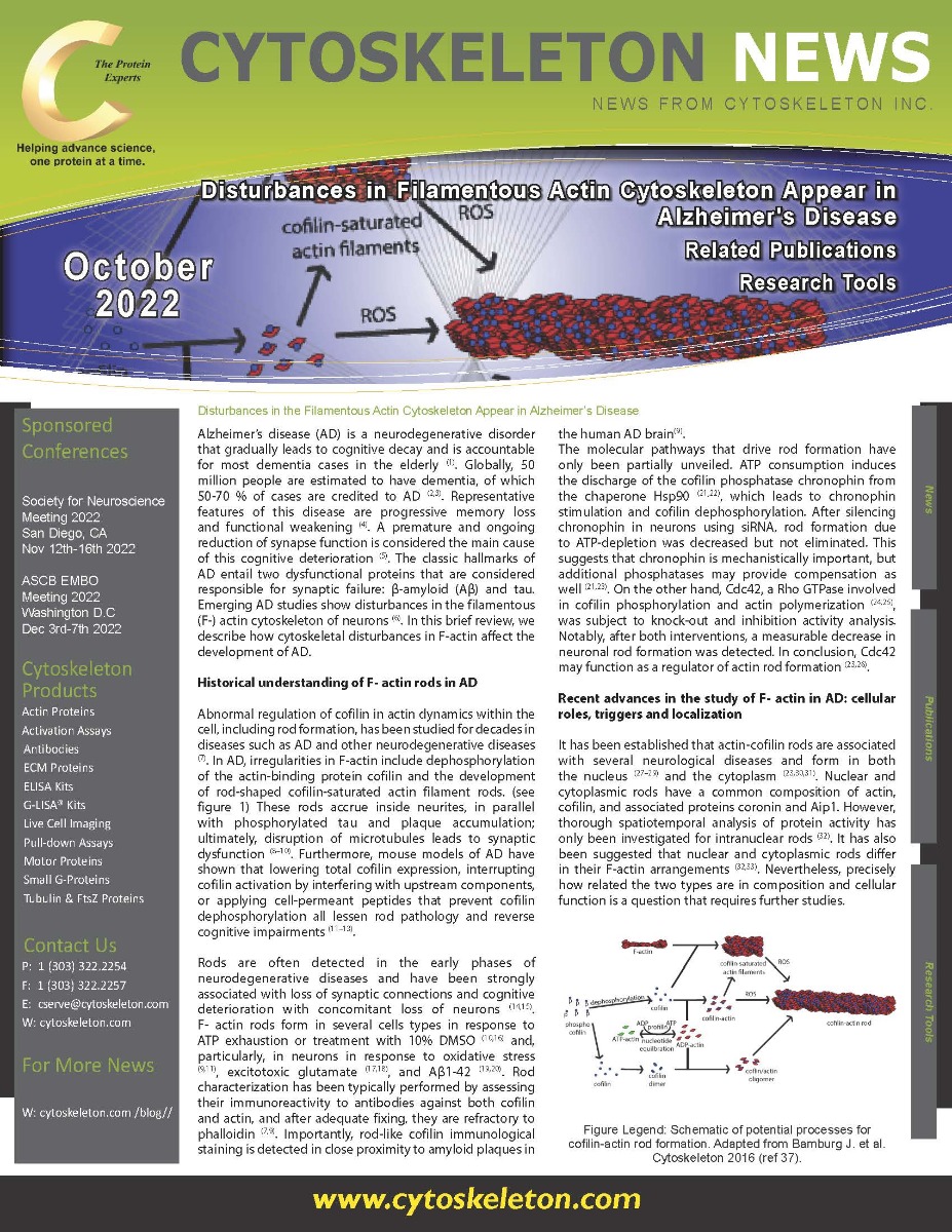
.")
Figure Legend: Schematic of potential processes for cofilin-actin rod formation. Adapted from Bamburg J. et al. Cytoskeleton 2016 (ref 37).
Recent advances in the study of F‐ actin in AD: cellular roles, triggers and localization
It has been established that actin-cofilin rods are associated with several neurological diseases and form in both the nucleus (27–29) and the cytoplasm (23,30,31). Nuclear and cytoplasmic rods have a common composition of actin, cofilin, and associated proteins coronin and Aip1. However, thorough spatiotemporal analysis of protein activity has only been investigated for intranuclear rods (32). It has also been suggested that nuclear and cytoplasmic rods differ in their F-actin arrangements (32,33). Nevertheless, precisely how related the two types are in composition and cellular function is a question that requires further studies.
Regulation of actin structures through actin dynamics is essential in many critical cell functions. Polymerization of actin is an energy-demanding process that can consume a significant portion of the available cellular ATP (7,34). A current, working hypothesis is that actin-cofilin rods appear as a cellular strategy to lower energy intake by discontinuing steady actin turnover during times of specific cell stress. This idea is supported by findings that metabolism and actin remodeling are interdependent processes (35). Furthermore, Ishikawa-Ankerhold et al. (7) designed a model to analyze cytoplasmic actin rod assembly in response to energy modulation. They discovered that cytoplasmic acidification promotes the accumulation of actin-cofilin rods, which implies that a local decrease in pH could be the source of cytoplasmic rod materialization.
Conclusions
Actin processes hold a critical role in the regulation of basic cell functions. Apart from AD, numerous neurological degenerative disorders, such as Parkinson’s, Huntington’s, and fronto-temporal dementia are also known for the accumulation of protein deposits in particular areas of the brain (36). In particular, abnormalities in cerebral F-actin in the shape of cofilin-actin rods have also been detected in Down syndrome (8). These cytoskeletal-related irregularities could contribute to dendritic spine malfunction and other pathological features, including neurofibrillary tangles (8). However, more studies are needed to better understand the mechanisms that underlie rod accumulation in the cytoplasmic or nuclear space, how this accumulation is regulated by physiological/pathological factors, and whether therapeutic intervention to reverse rod formation is sufficient to treat AD.
References
- Scheltens P, Blennow K, Breteler MMB, de Strooper B, Frisoni GB, Salloway S, et al. Alzheimer’s disease. Lancet. 2016 Jul 30;388(10043):505–17.
- Nichols E, Szoeke CEI, Vollset SE, Abbasi N, Abd-Allah F, Abdela J, et al. Global, regional, and national burden of Alzheimer’s disease and other dementias, 1990–2016: a systematic analysis for the Global Burden of Disease Study 2016. Lancet Neurol. 2019 Jan 1;18(1):88–106.
- Niu H, Álvarez-Álvarez I, Guillén-Grima F, Aguinaga-Ontoso I. Prevalencia e incidencia de la enfermedad de Alzheimer en Europa: metaanálisis. Neurologia. 2017 Oct 1;32(8):523–32.
- Mokdad A, Ballestros K, Echko M, Glenn S, Jama HO-, 2018 undefined. The state of US health, 1990-2016: burden of diseases, injuries, and risk factors among US states. jamanetwork.com [Internet]. 2018 Apr 10 [cited 2022 Jul 5];319(14):1444–72. Available from: https://jamanetwork.com/journals/jama/article-abstract/2678018
- Arendt T. Synaptic degeneration in Alzheimer’s disease. Acta Neuropathol [Internet]. 2009 [cited 2022 Jul 5];118(1):167–79. Available from: https://pubmed.ncbi.nlm.nih.gov/19390859/
- Bamburg JR, Minamide LS, Wiggan O, Tahtamouni LH, Kuhn TB. Cofilin and Actin Dynamics: Multiple Modes of Regulation and Their Impacts in Neuronal Development and Degeneration. Cells [Internet]. 2021 Oct 1 [cited 2022 Aug 5];10(10). Available from: https://pubmed.ncbi.nlm.nih.gov/34685706/
- Ishikawa-Ankerhold HC, Kurzbach S, Kinali AS, Müller-Taubenberger A. Formation of Cytoplasmic Actin-Cofilin Rods is Triggered by Metabolic Stress and Changes in Cellular pH. Front cell Dev Biol [Internet]. 2021 Nov 17 [cited 2022 Jul 21];9. Available from: https://pubmed.ncbi.nlm.nih.gov/34869330/
- Bamburg JR, Bernstein BW, Davis RC, Flynn KC, Goldsbury C, Jensen JR, et al. ADF/cofilin-actin rods in neurodegenerative diseases. Curr Alzheimer Res [Internet]. 2010 Apr 3 [cited 2022 Jul 18];7(3):241. Available from: /pmc/articles/PMC4458070/
- Minamide LS, Striegl AM, Boyle JA, Meberg PJ, Bamburg JR. Neurodegenerative stimuli induce persistent ADF/cofilin-actin rods that disrupt distal neurite function. Nat Cell Biol [Internet]. 2000 Sep [cited 2022 Jul 18];2(9):628–36. Available from: https://pubmed.ncbi.nlm.nih.gov/10980704/
- Minamide LS, Maiti S, Boyle JA, Davis RC, Coppinger JA, Bao Y, et al. Isolation and characterization of cytoplasmic cofilin-actin rods. J Biol Chem [Internet]. 2010 Feb 19 [cited 2022 Jul 18];285(8):5450–60. Available from: http://www.jbc.org/article/S0021925819375234/fulltext
- Woo JA, Boggess T, Uhlar C, Wang X, Khan H, Cappos G, et al. RanBP9 at the intersection between cofilin and Aβ pathologies: rescue of neurodegenerative changes by RanBP9 reduction. Cell Death Dis [Internet]. 2015 Mar 5 [cited 2022 Aug 5];6(3):1676. Available from: /pmc/articles/PMC4385917/
- Woo JA, Zhao X, Khan H, Penn C, Wang X, Joly-Amado A, et al. Slingshot-Cofilin activation mediates mitochondrial and synaptic dysfunction via Aβ ligation to β1-integrin conformers. Cell Death Differ [Internet]. 2015 Jun 1 [cited 2022 Aug 5];22(6):921–34. Available from: https://pubmed.ncbi.nlm.nih.gov/25698445/
- Deng Y, Wei J, Cheng J, Zhong P, Xiong Z, Liu A, et al. Partial Amelioration of Synaptic and Cognitive Deficits by Inhibiting Cofilin Dephosphorylation in an Animal Model of Alzheimer’s Disease. J Alzheimers Dis [Internet]. 2016 [cited 2022 Aug 5];53(4):1419–32. Available from: https://pubmed.ncbi.nlm.nih.gov/27372643/
- Maloney MT, Bamburg JR. Cofilin-mediated neurodegeneration in Alzheimer’s disease and other amyloidopathies. Mol Neurobiol [Internet]. 2007 [cited 2022 Jul 20];35(1):21–43. Available from: https://pubmed.ncbi.nlm.nih.gov/17519504/
- Shu L, Chen B, Chen B, Xu H, Wang G, Huang Y, et al. Brain ischemic insult induces cofilin rod formation leading to synaptic dysfunction in neurons. J Cereb Blood Flow Metab [Internet]. 2019 Nov 1 [cited 2022 Aug 5];39(11):2181. Available from: /pmc/articles/PMC6827117/
- Fukui Y, Katsumaru H. Nuclear actin bundles in Amoeba, Dictyostelium and human HeLa cells induced by dimethyl sulfoxide. Exp Cell Res [Internet]. 1979 [cited 2022 Jul 20];120(2):451–5. Available from: https://pubmed.ncbi.nlm.nih.gov/571346/
- Davis RC, Maloney MT, Minamide LS, Flynn KC, Stonebraker MA, Bamburg JR. Mapping cofilin-actin rods in stressed hippocampal slices and the role of cdc42 in amyloid-beta-induced rods. J Alzheimers Dis [Internet]. 2009 [cited 2022 Aug 5];18(1):35–50. Available from: https://pubmed.ncbi.nlm.nih.gov/19542631/
- Bie B, Wu J, Foss JF, Naguib M. Amyloid fibrils induce dysfunction of hippocampal glutamatergic silent synapses. Hippocampus [Internet]. 2018 Aug 1 [cited 2022 Aug 5];28(8):549–56. Available from: https://pubmed.ncbi.nlm.nih.gov/29704282/
- Maloney MT, Minamide LS, Kinley AW, Boyle JA, Bamburg JR. Beta-secretase-cleaved amyloid precursor protein accumulates at actin inclusions induced in neurons by stress or amyloid beta: a feedforward mechanism for Alzheimer’s disease. J Neurosci [Internet]. 2005 Dec 7 [cited 2022 Jul 20];25(49):11313–21. Available from: https://pubmed.ncbi.nlm.nih.gov/16339026/
- Kang DE, Woo JA. Cofilin, a Master Node Regulating Cytoskeletal Pathogenesis in Alzheimer’s Disease. J Alzheimers Dis [Internet]. 2019 [cited 2022 Aug 5];72(s1):S131–44. Available from: https://pubmed.ncbi.nlm.nih.gov/31594228/
- Huang TY, Minamide LS, Bamburg JR, Bokoch GM. Chronophin Mediates an ATP-Sensing Mechanism for Cofilin Dephosphorylation and Neuronal Cofilin-Actin Rod Formation. Dev Cell. 2008 Nov 11;15(5):691–703.
- Aragona M, Panciera T, Manfrin A, Giulitti S, Michielin F, Elvassore N, et al. A mechanical checkpoint controls multicellular growth through YAP/TAZ regulation by actin-processing factors. Cell [Internet]. 2013 Aug 29 [cited 2022 Aug 5];154(5):1047–59. Available from: http://www.cell.com/article/S0092867413009513/fulltext
- Walter LM, Rademacher S, Pich A, Claus P. Profilin2 regulates actin rod assembly in neuronal cells. Sci Reports 2021 111 [Internet]. 2021 May 13; 11(1):1–13. Available from: https://www.nature.com/articles/s41598-021-89397-9
- Dubreuil CI, Van Vactor DL, Van Vactor DL. Signal Transduction Pathways: From Receptor to the Actin Cytoskeleton. 2011 [cited 2022 Jul 19];235–63. Available from: https://link.springer.com/chapter/10.1007/978-1-4419-7368-9_12
- Zhao ZS, Manser E. PAK and other Rho-associated kinases--effectors with surprisingly diverse mechanisms of regulation. Biochem J [Internet]. 2005 Mar 1; 386(Pt 2):201–14. Available from: https://pubmed.ncbi.nlm.nih.gov/15548136/
- Bamburg JR, Bloom GS. Cytoskeletal pathologies of Alzheimer disease. Cell Motil Cytoskeleton [Internet]. 2009 Aug [cited 2022 Jul 18];66(8):635–49. Available from: https://pubmed.ncbi.nlm.nih.gov/19479823/
- Kloc M, Chanana P, Vaughn N, Uosef A, Kubiak JZ, Ghobrial RM. New Insights into Cellular Functions of Nuclear Actin. Biol 2021, Vol 10, Page 304 [Internet]. 2021 Apr 7 [cited 2022 Aug 4];10(4):304. Available from: https://www.mdpi.com/2079-7737/10/4/304/htm
- Biel N, Figard L, Sokac AM. Imaging Intranuclear Actin Rods in Live Heat Stressed Drosophila Embryos. JoVE (Journal Vis Exp [Internet]. 2020 May 15 [cited 2022 Aug 4];2020(159):e61297. Available from: https://www.jove.com/es/v/61297/imaging-intranuclear-actin-rods-live-heat-stressed-drosophila
- Munsie LN, Desmond CR, Truant R. Cofilin nuclear-cytoplasmic shuttling affects cofilin-actin rod formation during stress. J Cell Sci [Internet]. 2012 Sep 1 [cited 2022 Aug 4];125(17):3977–88. Available from: https://journals.biologists.com/jcs/article/125/17/3977/32560/Cofilin-nuclear-cytoplasmic-shuttling-affects
- Wang Q, Yuan W, Yang X, Wang Y, Li Y, Qiao H. Role of Cofilin in Alzheimer’s Disease. Front Cell Dev Biol [Internet]. 2020 Nov 26 [cited 2022 Aug 4];8:584898. Available from: /pmc/articles/PMC7726191/
- Schonhofen P, Medeiros LM, Chatain C, Bristot I, Klamt F. Cofilin/actin rod formation by dysregulation of cofilin-1 activity as a central initial step in neurodegeneration. Mini Rev Med Chem [Internet]. 2014 May 16 [cited 2022 Aug 4];14(5):393–400. Available from: https://pubmed.ncbi.nlm.nih.gov/24813767/
- Ishikawa-Ankerhold HC, Daszkiewicz W, Schleicher M, Müller-Taubenberger A. Actin-Interacting Protein 1 Contributes to Intranuclear Rod Assembly in Dictyostelium discoideum. Sci Reports 2017 71 [Internet]. 2017 Jan 11 [cited 2022 Aug 4];7(1):1–12. Available from: https://www.nature.com/articles/srep40310
- Aizawa H, Katadae M, Maruya M, Sameshima M, Murakami-Murofushi K, Yahara I. Hyperosmotic stress-induced reorganization of actin bundles in Dictyostelium cells over-expressing cofilin. Genes Cells [Internet]. 1999 [cited 2022 Aug 4];4(6):311–24. Available from: https://pubmed.ncbi.nlm.nih.gov/10421841/
- Bernstein BW, Bamburg JR. Actin-ATP hydrolysis is a major energy drain for neurons. J Neurosci [Internet]. 2003 Jan 1 [cited 2022 Jul 21];23(1):1–6. Available from: https://pubmed.ncbi.nlm.nih.gov/12514193/
- DeWane G, Salvi AM, DeMali KA. Fueling the cytoskeleton-links between cell metabolism and actin remodeling. J Cell Sci. 2021 Feb 1;134(3).
- Lee SJ, Lim HS, Masliah E, Lee HJ. Protein aggregate spreading in neurodegenerative diseases: problems and perspectives. Neurosci Res [Internet]. 2011 Aug [cited 2022 Jul 18];70(4):339–48. Available from: https://pubmed.ncbi.nlm.nih.gov/21624403/
- Bamburg, J.R. and Bernstein, B.W. (2016), Actin dynamics and cofilin-actin rods in alzheimer disease. Cytoskeleton, 73: 477-497. https://doi.org/10.1002/cm.21282