Exoenzyme C3 transferase protein: His tagged:Clostridium botulinum recombinant

Exoenzyme C3 transferase protein: His tagged:Clostridium botulinum recombinant
$0.00

Product Uses Include

  • Inhibition of RhoA, B and C in vitro
  • Inhibition of RhoA, B and C in vivo (See Table 1 for examples of use)

Material
Exoenzyme C3 transferase is an ADP ribosyl transferase that selectively ribosylates RhoA, RhoB and RhoC proteins on asparagine residue 41, rendering them inactive. It has extremely low affinity for other members of the Rho family such as Cdc42 and Rac1 and does therefore not affect these GTPases. Hence, C3 transferase is a very potent and useful reagent to specifically block RhoA/B/C signaling.

C3 transferase has been produc ed by expression in E. coli as a His-tagged protein. The recombinant protein is 24 kDa in size and is supplied as a lyophilized powder. Reconstitution of the protein in water to 1 mg/ml leaves the protein in the following buffer: 20 mM Tris pH 7.5, 50 mM NaCl, 0.5% sucrose and 0.1% dextran. Protein concentration is determined by the Precision Red Advanced Protein Assay Reagent Cat. # ADV02.

C3 transferase protein is also available in a cell permeable format (Cat. CT04) for fast, efficient and simple inhibition of RhoA/B/C in living cells.
 

Purity
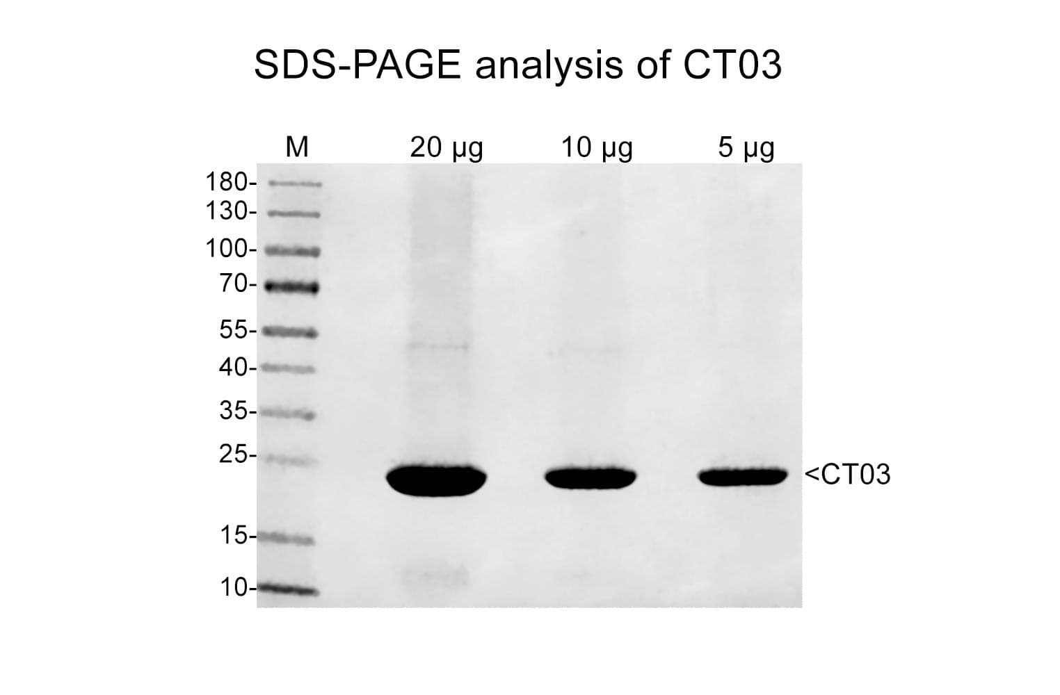
Purity is determined by scanning densitometry of protein run on SDS-PAGE gels. CT03 consists of more than 90% pure exoenzyme C3 Transferase.

ct03gel

Figure 1: Exoenzyme C3 transferase purity determination.  A 20 µg sample of CT03 was separated by SDS-PAGE and protein was stained with coomassie blue.  Protein quantitation was performed using Precision Red Assay reagent (Cat. # ADV02).  Purity was determined by scanning densitometry. The protein was determined to be >90% pure.

Biological Activity
Biological activity of C3 transferase is verified by the ability of the protein to ribosylate RhoA protein in platelet lysates in vitro (Fig. 2).

ct03qcblot

Figure 1: ADP-ribosylation of RhoA protein in human platelet extract.  Platelet extract (100 µg) was reacted with C3 protein (1 µg) for 30 min at 37°C.  Extracts were run on  non-denaturing gel electrophoresis and RhoA protein was detected by Western blot.  Lane 1 shows untreated extracts.  Lane 2 shows C3 transferase treated extracts.  The RhoA in the C3 transferase treated extract shows increased migration in the gel due to its ADP ribosylation. 

Table 1. Examples of how Cytoskeleton, Inc's C3 transferase has been used on cells to inactivate RhoA.

Cell type

Method of introduction into cells

Concentration of C3 used (mg/ml)

Citation

BS-C-1

microinjection

0.1

2

NRK

added to culture media

0.02

7

Xenopus oocytes

microinjection

0.08

1

Primary Aplysia bag cells

microinjection

0.3

8

For product Datasheets and MSDSs please click on the PDF links below.   For additional information, click on the FAQs tab above or contact our Technical Support department at tservice@cytoskeleton.com

AuthorTitleJournalYearArticle Link
Sun, Xiaojing et al.Chemerin Enhances Migration and Invasion of OC Cells via CMKLR1/RhoA/ROCK-Mediated EMTInternational Journal of Endocrinology2024
Ciszewski, Wojciech M. et al.Diverse roles of SARS-CoV-2 Spike and Nucleocapsid proteins in EndMT stimulation through the TGF-β-MRTF axis inhibited by aspirinCell Communication and Signaling2024
Ciszewski, Wojciech M. et al.SARS-CoV-2 S and N protein peptides drive invasion abilities of colon cancer cells through TGF-β1 regulationBiochimica et Biophysica Acta (BBA) - Molecular Cell Research2023
Wang, Tianshi et al.Regulation of the Hippo/YAP axis by CXCR7 in the tumorigenesis of gastric cancerJournal of Experimental and Clinical Cancer Research2023
Hui, Justin et al.Coordinated efforts of different actin filament populations are needed for optimal cell wound repairMolecular Biology of the Cell2023
do Rosário, Carline Fermino et al.Lateral and longitudinal compaction of PRC1 overlap zones drives stabilization of interzonal microtubulesMolecular Biology of the Cell2023
Harasimov, Katarina et al.Actin-driven chromosome clustering facilitates fast and complete chromosome capture in mammalian oocytesNature Cell Biology2023
Ciszewski, Wojciech M. et al.SARS-CoV-2 S and N protein peptides drive invasion abilities of colon cancer cells through TGF-β1 regulationBiochimica et Biophysica Acta (BBA) - Molecular Cell Research2023
Zhang, Xingyu et al.Soluble TREM2 ameliorates tau phosphorylation and cognitive deficits through activating transgelin-2 in Alzheimer’s diseaseNature Communications2023
Harasimov, Katarina et al.Actin-driven chromosome clustering facilitates fast and complete chromosome capture in mammalian oocytesNature Cell Biology 20232023
Guo, Yan et al.Opposing roles of ZEB1 in the cytoplasm and nucleus control cytoskeletal assembly and YAP1 activityCell Reports2022
Du, Xing et al.The mevalonate pathway promotes the metastasis of osteosarcoma by regulating YAP1 activity via RhoAGenes & Diseases2022
Xie, Lang et al.MYO1B enhances colorectal cancer metastasis by promoting the F-actin rearrangement and focal adhesion assembly via RhoA/ROCK/FAK signalingAnnals of Translational Medicine2021
Zhou, Qun et al.Inflammatory Immune Cytokine TNF-α Modulates Ezrin Protein Activation via FAK/RhoA Signaling Pathway in PMVECs HyperpermeabilityFrontiers in Pharmacology2021
Landino, Jennifer et al.Rho and F-actin self-organize within an artificial cell cortexCurrent Biology2021
McCray, Brett A. et al.Neuropathy-causing TRPV4 mutations disrupt TRPV4-RhoA interactions and impair neurite extensionNature Communications2021
Pal, Debadrita et al.Rac and Arp2/3-Nucleated Actin Networks Antagonize Rho During Mitotic and Meiotic CleavagesFrontiers in Cell and Developmental Biology2020
Lu, Jiaoyang et al.Basement Membrane Regulates Fibronectin Organization Using Sliding Focal Adhesions Driven by a Contractile WinchDevelopmental Cell2020
Wang, Lei et al.YAP and TAZ protect against white adipocyte cell death during obesityNature Communications2020
Evans, Frances et al.Signaling pathways in cytoskeletal responses to plasma membrane depolarization in corneal endothelial cellsJournal of Cellular Physiology2020
Miao, Hui et al.Cell ratcheting through the Sbf RabGEF directs force balancing and stepped apical constrictionJournal of Cell Biology2019
Ko, Clint S. et al.Microtubules promote intercellular contractile force transmission during tissue foldingJournal of Cell Biology2019
Zahra, Fatema Tuz et al.Endothelial RhoA GTPase is essential for in vitro endothelial functions but dispensable for physiological in vivo angiogenesisScientific Reports2019
Hübner, Kathleen et al.Wnt/β-catenin signaling regulates VE-cadherin-mediated anastomosis of brain capillaries by counteracting S1pr1 signalingNature Communications2018
Song, Nan et al.NLRP3 Phosphorylation Is an Essential Priming Event for Inflammasome ActivationMolecular Cell2017
Ren, Yu et al.Induction of cell cycle arrest by increasing GTP-RhoA levels via taxol-induced microtubule polymerization in renal cell carcinomaMolecular Medicine Reports2017
Platet, Nadine et al.The tumor suppressor CDX2 opposes pro-metastatic biomechanical modifications of colon cancer cells through organization of the actin cytoskeletonCancer Letters2017
Raya-Sandino, Arturo et al.Zonula occludens-2 regulates Rho proteins activity and the development of epithelial cytoarchitecture and barrier functionBiochimica et Biophysica Acta - Molecular Cell Research2017
Mason, Frank M. et al.RhoA GTPase inhibition organizes contraction during epithelial morphogenesisJournal of Cell Biology2016
Park, Yong Hwan et al.Pyrin inflammasome activation and RhoA signaling in the autoinflammatory diseases FMF and HIDSNature Immunology2016
Tivari, Samir et al.An in vitro dormancy model of estrogen-sensitive breast cancer in the bone marrow: A tool for molecular mechanism studies and hypothesis generationJournal of Visualized Experiments2015
Lemons, M L et al.Integrins and cAMP mediate netrin-induced growth cone collapseBrain Research2013
Takefuji, Mikito et al.RhoGEF12 controls cardiac remodeling by integrating G protein- and integrindependent signaling cascadesJournal of Experimental Medicine2013
Wong, Hon Kit et al.Merlin/NF2 regulates angiogenesis in schwannomas through a Rac1/semaphorin 3F-dependent mechanismNeoplasia2012
Takefuji, Mikito et al.G13-mediated signaling pathway is required for pressure overload-induced cardiac remodeling and heart failureCirculation2012
Melendez, Jaime et al.RhoA GTPase Is Dispensable for Actomyosin Regulation but Is Essential for Mitosis in Primary Mouse Embryonic Fibroblasts * □ S2011
Toullec, Aurore et al.Oxidative stress promotes myofibroblast differentiation and tumour spreadingEMBO Molecular Medicine2010
Marshall, Andrew K. et al.ERK1/2 signaling dominates over RhoA signaling in regulating early changes in RNA expression induced by endothelin-1 in neonatal rat cardiomyocytesPLoS ONE2010
Oblander, Samantha A. et al.Distinct PTPmu-associated signaling molecules differentially regulate neurite outgrowth on E-, N-, and R-cadherinMolecular and Cellular Neuroscience2010
Benink, Hélène A. et al.Concentric zones of active RhoA and Cdc42 around single cell woundsJournal of Cell Biology2005
O'neil, Caroline H et al.Stimulation of Vascular Smooth Muscle Cell Proliferation and Migration by Apolipoprotein(a) Is Dependent on Inhibition of Transforming Growth Factor-β Activation and on the Presence of Kringle IV Type 9*2004
Ridley, Anne J.Pulling Back to Move ForwardCell2004
Simpson, Kaylene J. et al.Functional analysis of the contribution of RhoA and RhoC GTPases to invasive breast carcinomaCancer Research2004
Burakov, Anton et al.Centrosome positioning in interphase cellsJournal of Cell Biology2003
Valderrama, Ferran et al.The Golgi-associated COPI-coated buds and vesicles contain β/γ-actinProceedings of the National Academy of Sciences of the United States of America2000

Question 1:  What is the difference between the Rho inhibitors CT03 and CT04?

Answer 1:  The only difference between these C3 Transferase proteins (Cat. # CT03 and CT04) is that CT04 is covalently linked to a proprietary cell penetrating moiety via a disulfide bond.  In this way, CT04 is a much better reagent to use to inhibit Rho activity in living cells.


Question 2:  How can I assess whether Rho activity is changing following CT03 treatment? 

Answer 2:  There are multiple ways to measure changes in Rho activity.  If CT03 has been delivered to cells via micro-injection or pinocytic uptake, we recommend examining Rho-mediated stress fiber formation with fluorescently-labeled phalloidin (Cat. # PHDG1, PHDH1, PHDN1, PHDR1).  These Acti-stain phalloidins label F-actin stress fibers.  Activation of Rho can be directly quantified with one of our activation assays, either the traditional pull-down (Cat. # BK036) or the RhoA G-LISA activation assay (Cat. # BK124).  You can also measure CT03’s ability to ADP ribosylate native Rho in human platelet extracts in vitro (please see the CT03 datasheet for more details).  Briefly, a standard biological assay for monitoring the ADP ribosylation of Rho consists of an in vivo ribosylation reaction followed by non-denaturing gel electophoresis and Western blot analysis using Cytoskeleton’s anti-Rho monoclonal antibody (Cat. # ARH03).  Stringent quality control ensures that >80% of native Rho protein is ADP ribosylated by the recombinant C3 transferase.


If you have any questions concerning this product, please contact our Technical Service department at tservice@cytoskeleton.com.