* Limited stock available. If stock is not available, Cytoskeleton will produce a new batch upon request. Minimum order will apply. Inquire for more information.
Product Uses
- Study of K-Ras4B exchange activity with different GEFs.
- Identification of K-Ras4B exchange factors (GEFs)
- Positive control for GEF studies.
- Biochemical characterization of K-Ras4B protein interactions
- Western blot standard
Materials
The wild-type human K-Ras4B protein has been produced in a bacterial expression system. The recombinant protein contains six histidine residues at its amino terminus (His-tag). The molecular weight of 6xHis tagged K-Ras4B is approximately 25 kDa and is supplied as a white lyophilized powder.
Storage
Before reconstitution, briefly centrifuge to collect the product at the bottom of the tube. The protein should be reconstituted to 5 mg/ml with the addition of 20 µl of Milli-Q water (100 µg size). When reconstituted, the protein will be in the following buffer: 50 mM Tris pH 7.5, 50 mM NaCl, 0.5 mM MgCl2, 5% (w/v) sucrose and 1% (w/v) dextran. In order to maintain high biological activity of the protein it is strongly recommended that the protein solution be supplemented with DTT to 1 mM final concentration, aliquoted into "experiment sized" amounts, snap frozen in liquid nitrogen and stored a -70°C. The protein is stable for six months if stored at -70°C. The protein should not be exposed to repeated freeze-thaw cycles. The lyophilized protein is stable at 4°C desiccated (<10% humidity) for one year.
Purity
Protein purity is determined by scanning densitometry of Coomassie Blue stained protein on a 4-20% polyacrylamide gradient gel. His tagged K-Ras4B protein was determined to be >90% pure. (see Figure 1).
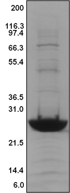
Legend: A 20 µg sample of recombinant K-Ras4B protein (molecular weight approx. 25 kDa) was separated by electrophoresis in a 4-20% SDS-PAGE system and stained with Coomassie Blue. Protein quantitation was determined using the Precision Red Protein Assay Reagent (Cat. # ADV02). Mark12 molecular weight markers are from Life Technologies Inc.
Biological Activity Assay
The biological activity of K-Ras4B can be determined from the ability of the SOS1 exchange domain (SOS1-ExD) to catalyze the exchange of GDP for GTP on K-Ras4B. A standard biological assay for monitoring the biological activity of K-Ras4B is an exchange assay utilizing the 2X Exchange Buffer from the Rho-GEF exchange assay kit (Cat.# BK100) and the human SOS1 GEF domain (Cat.# CS-SOS1).
Reagents
1. Recombinant K-Ras4B protein (Cat.# CS-RS03)
2. Recombinant SOS1-ExD protein (Cat.# CS-SOS1)
3. 2x Exchange Buffer (40 mM Tris pH 7.5, 100 mM NaCl, 20 mM MgCl2 , 0.1 mg/ml BSA, 1.5 µM mant-GTP)
4. Dilution Buffer (20 mM Tris pH 7.5, 50 mM NaCl, 10 mM MgCl2, 0.1 mg/ml BSA)
Equipment
1. Fluorescence spectrometer (λex=360nm, λem=440nm)
2. Corning 96-well half area plates (Cat # 3686) or other plate with low protein binding surface.
Method
1. Dilute SOS1-ExD protein (Cat# CS-SOS1) to 1 µM (0.06 mg/ml) with Dilution Buffer.
2. Dilute K-Ras4B to 50 µM (1.1 mg/ml) with Dilution Buffer.
3. Dissolve lyophilized 2x Exchange Buffer in 5 ml Milli-Q water and keep in room temperature.
4. Set up the plate reader for kinetic fluorescence measurements (Excitation wavelength at 360 nm and emission wavelength at 440 nm) with readings every 30 seconds for 30 minutes.
5. Add the following components together and mix well by gentle pipetting:
Exchange reaction mix / 96 well black plate
2x Exchange Buffer / 50 μl
dH2O / 26 μl
50 μM K-Ras4B / 4 μl
6. Pipette 20 µl of 1 µM SOS1-ExD protein or Dilution Buffer in their respective wells and immediately pipette up and down twice and begin reading the fluorescence.
7. Once the readings are complete and the plate reader file has been saved, the exchange rate can be calculated by reducing the data to Vmax with the software that accompanies the plate reader.
Figure 2. SOS1-ExD protein mediated mant-GTP exchange on K-Ras4B

Figure 1. K-Ras4B Protein Purity Determination
Legend: K-Ras4B protein was added to the wells of a 96-well half area plate containing diluted Exchange Buffer and mixed well. To initiate the exchange reaction, SOS1-ExD protein (red triangles), or Dilution Buffer (grey triangles), was added to the wells, mixed, and fluorescence measurements were obtained using a Tecan SpectraFluor Plus Spectrophotometer. The average fluorescence data from quadruplicate assays were normalized to the fluorescence at the zero time point. The resulting data were plotted as the change in relative fluorescence units (DRFU) over time using GraphPad Prism software.
For product Datasheets and MSDSs please click on the PDF links below.
If you have any questions concerning this product, please contact our Technical Service department at tservice@cytoskeleton.com
Coming soon! If you have any questions concerning this product, please contact our Technical Service department at tservice@cytoskeleton.com


