Tubulin protein (fluorescent HiLyte 647): porcine brain

Tubulin protein (fluorescent HiLyte 647): porcine brain
$0.00

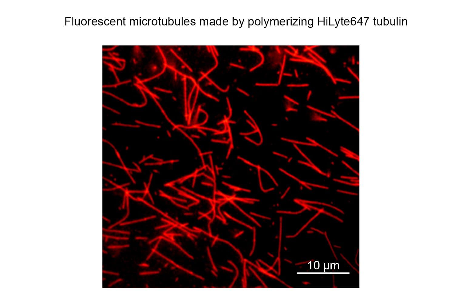
HiLyte Fluor™ 647labeled microtubules formed from HiLyte Fluor™ 647 labeled tubulin.

TL670MMTs
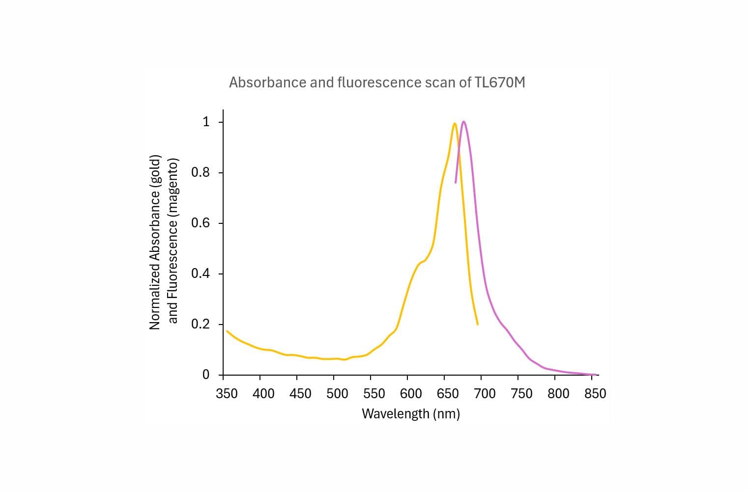
TL670_scan

Product Uses Include

  • Laser based applications
  • Monitoring microtubule dynamcs in living cells
  • Speckle microscopy
  • Formation of fluorescent microtubules
  • Microscopy studies of MAP and microtubule associated motor activities
  • Nanotechnology

Material
Porcine brain tubulin (>99% pure, see Cat. # T240) has been modified to contain covalently linked HiLyte Fluor™ 647 (HiLyte Fluor is a trademark of Anaspec Inc, CA) at random surface lysines. An activated ester of HiLyte Fluor™ 647 was used to label the protein. Labeling stoichiometry was determined by spectroscopic measurement of protein and dye concentrations (dye extinction coefficient when protein bound is 250,000M-1cm-1). Final labeling stoichiometry is 0.2 to 0.7 dyes per tubulin heterodimer. HiLyte Fluor™ 647 labeled tubulin can be detected using a filter set of 600-630 nm excitation and 660-680 emission. HiLyte Fluor™ 647 tubulin is in a versatile, stable and easily shipped format. It is ready for micro-injection or in vitro polymerization. Cytoskeleton, Inc. also offers AMCA (Cat. # TL440M), HiLyte Fluor™ 488 (Cat. # TL488M), rhodamine (Cat. # TL590M), X-rhodamine (Cat. # TL620M) labeled tubulins.


Purity
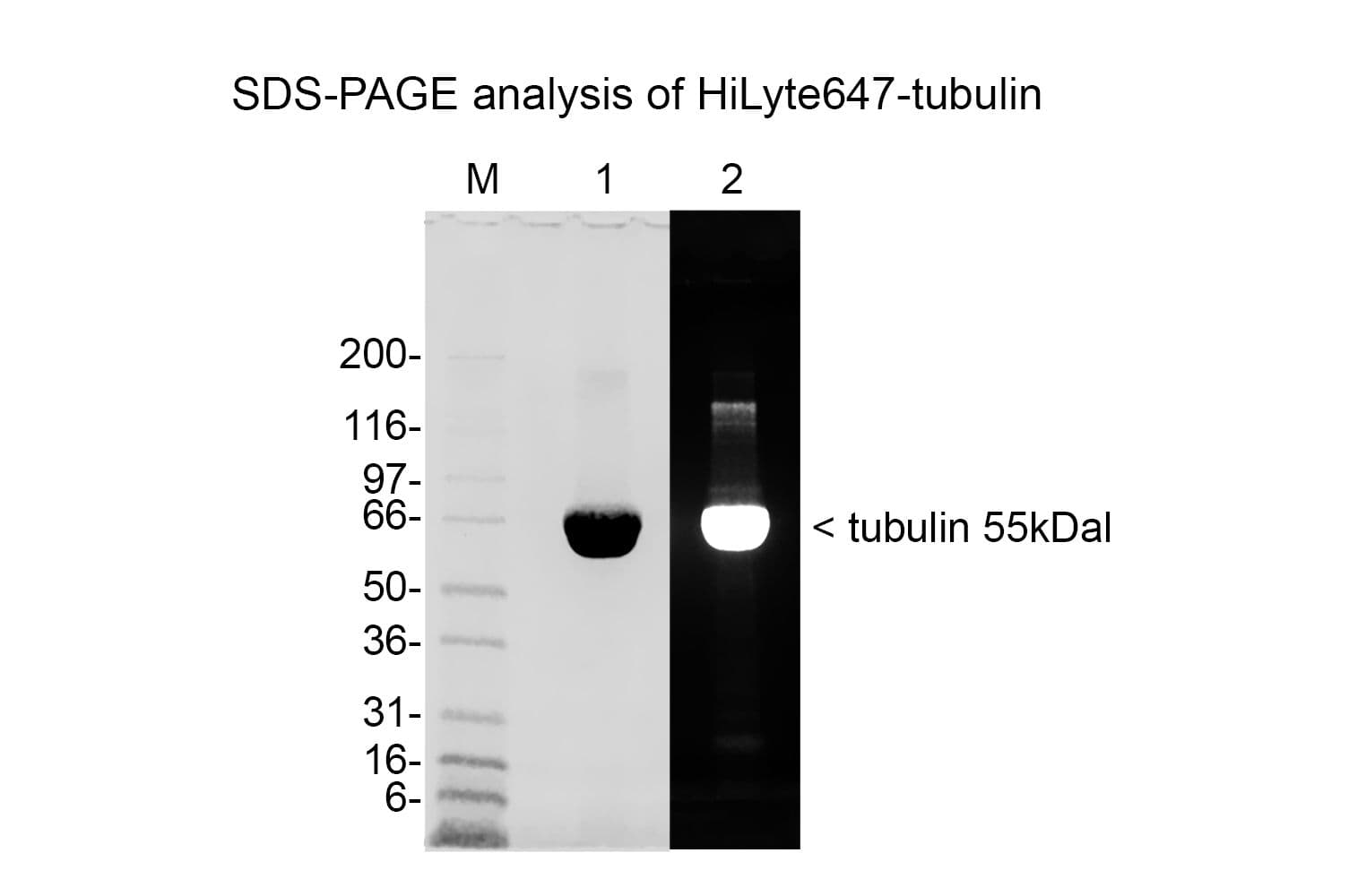
The protein purity of the tubulin used for labeling is determined by scanning densitometry of Coomassie Blue stained protein on a 4-20% polyacrylamide gel. The protein used for TL670M is >99% pure tubulin (Figure 1 A). Labeled protein is run on an SDS gel and photographed under UV light. Any unincorporated HiLyte Fluor™ 670
 dye would be visible in the dye front. No fluorescence is detected in the dye front, indicating that no free dye is present in the final product (Figure 1 B).

tl334mgels

Figure 1: HiLyte Fluor™ 647 tubulin protein purity determination. A 50 µg sample of unlabeled tubulin protein was separated by electrophoresis in a 4-20% SDS-PAGE system and stained with Coomassie Blue (A). Protein quantitation was performed using the Precision Red Protein Assay Reagent (Cat. # ADV02). 20 µg of the same protein sample was run in a 4-20% SDS-PAGE system and photographed directly under 525-625nm illumination (B).

Biological Activity
The biological activity of HiLyte Fluor™ 647 tubulin is assessed by a tubulin polymerization assay. To pass quality control, a 5 mg/ml solution of HiLyte Fluor™ 647 labeled tubulin in G-PEM plus 5% glycerol must polymerize to >85%. This is comparable to unlabeled tubulin under identical conditions.

For product Datasheets and MSDSs please click on the PDF links below.   For additional information, click on the FAQs tab above or contact our Technical Support department at tservice@cytoskeleton.com

AuthorTitleJournalYearArticle Link
Robinson, Benjamin P. et al.Septin-coated microtubules promote maturation of multivesicular bodies by inhibiting their motilityJournal of Cell Biology2024
Xu, Yixin et al.Partial closure of the γ-tubulin ring complex by CDK5RAP2 activates microtubule nucleationDevelopmental Cell2024
Takagishi, Maki et al.Motor protein Kif6 regulates cilia motility and polarity in brain ependymal cellsDMM Disease Models and Mechanisms2024
Seo, Dahee et al.Poxvirus A51R Proteins Negatively Regulate Microtubule-Dependent Transport by Kinesin-1International Journal of Molecular Sciences2024
Rai, Dipti et al.CAMSAPs and nucleation-promoting factors control microtubule release from γ-TuRCNature Cell Biology2024
Aquino-Perez, Cecilia et al.FAM110A promotes mitotic spindle formation by linking microtubules with actin cytoskeletonProceedings of the National Academy of Sciences2024
Nithianantham, Stanley et al.The kinesin-5 tail and bipolar minifilament domains are the origin of its microtubule crosslinking and sliding activityMolecular biology of the cell2023
McHugh, Toni et al.Potent microtubule-depolymerizing activity of a mitotic Kif18b–MCAK–EB networkJournal of Cell Science2023
Tsukita, Kazuto et al.Phase separation of an actin nucleator by junctional microtubules regulates epithelial functionScience Advances2023
Palumbo, Jacob et al.Directly Measuring Forces Within Reconstituted Active Microtubule BundlesJoVE (Journal of Visualized Experiments)2022
Havelka, Daniel et al.Lab-on-chip microscope platform for electro-manipulation of a dense microtubules networkScientific Reports2022
Tsitkov, Stanislav et al.The rate of microtubule breaking increases exponentially with curvatureScientific Reports 2022
Castrogiovanni, Cédric et al.Evidence for a HURP/EB free mixed-nucleotide zone in kinetochore-microtubulesNature Communications2022
Henty-Ridilla, Jessica L.Visualizing Actin and Microtubule Coupling Dynamics In Vitro by Total Internal Reflection Fluorescence (TIRF) MicroscopyJoVE (Journal of Visualized Experiments)2022
Planelles-Herrero, Vicente Jose et al.Elongator stabilizes microtubules to control central spindle asymmetry and polarized trafficking of cell fate determinantsNature Cell Biology 2022
Hochmair, Janine et al.Molecular crowding and RNA synergize to promote phase separation, microtubule interaction, and seeding of Tau condensatesThe EMBO Journal2022
Havelka, Daniel et al.Lab-on-chip microscope platform for electro-manipulation of a dense microtubules networkScientific Reports2022
Watson, Joseph L. et al.High-efficacy subcellular micropatterning of proteins using fibrinogen anchorsJournal of Cell Biology2021
Cheng, Xianrui et al.Xenopus laevis egg extract preparation and live imaging methods for visualizing dynamic cytoplasmic organizationJournal of Visualized Experiments2021
Saper, Gadiel et al.Robotic end-to-end fusion of microtubules powered by kinesinScience Robotics2021
Diwaker, Drishya et al.Deletion of the Pseudorabies virus gE/gI-US9p complex disrupts kinesin KIF1A and KIF5C recruitment during egress, and alters the properties of microtubule-dependent transport in vitroPLoS Pathogens2020
Aher, Amol et al.CLASP Mediates Microtubule Repair by Restricting Lattice Damage and Regulating Tubulin IncorporationCurrent Biology2020
Tseng, Kuo Fu et al.The Tail of Kinesin-14a in Giardia Is a Dual Regulator of MotilityCurrent Biology2020
Bassir Kazeruni, Neda M. et al.Microtubule Detachment in Gliding Motility Assays Limits the Performance of Kinesin-Driven Molecular ShuttlesLangmuir2020
Rodríguez-García, Ruddi et al.Mechanisms of Motor-Independent Membrane Remodeling Driven by Dynamic MicrotubulesCurrent Biology2020
Gaska, Ignas et al.The Mitotic Crosslinking Protein PRC1 Acts Like a Mechanical Dashpot to Resist Microtubule SlidingDevelopmental Cell2020
Zehr, Elena A. et al.Katanin Grips the β-Tubulin Tail through an Electropositive Double Spiral to Sever MicrotubulesDevelopmental Cell2020
von Appen, Alexander et al.LEM2 phase separation promotes ESCRT-mediated nuclear envelope reformationNature2020
Farhadi, Leila et al.Actin and microtubule crosslinkers tune mobility and control co-localization in a composite cytoskeletal networkSoft Matter2020
Nakos, Konstantinos et al.Regulation of microtubule plus end dynamics by septin 9Cytoskeleton2019
Guedes-Dias, Pedro et al.Kinesin-3 Responds to Local Microtubule Dynamics to Target Synaptic Cargo Delivery to the PresynapseCurrent Biology2019
Kazeruni, Neda M.Bassir et al.Assembling molecular shuttles powered by reversibly attached kinesinsJournal of Visualized Experiments2019
Siddiqui, Nida et al.PTPN21 and Hook3 relieve KIF1C autoinhibition and activate intracellular transportNature Communications2019
Jiang, Shuo et al.Interplay between the Kinesin and Tubulin Mechanochemical Cycles Underlies Microtubule Tip Tracking by the Non-motile Ciliary Kinesin Kif7Developmental Cell2019
Li, Feiran et al.Local direction change of surface gliding microtubulesBiotechnology and Bioengineering2019
Nakos, Konstantinos et al.Septin 2/6/7 complexes tune microtubule plus-end growth and EB1 binding in a concentration- And filament-dependent mannerMolecular Biology of the Cell2019
Karasmanis, Eva P. et al.Erratum: Polarity of Neuronal Membrane Traffic Requires Sorting of Kinesin Motor Cargo during Entry into Dendrites by a Microtubule-Associated Septin (Developmental Cell (2018) 46(2) (204–218.e7), (S1534580718304982) (10.1016/j.devcel.2018.06.013))Developmental Cell2018
Colin, Alexandra et al.Actin-Network Architecture Regulates Microtubule DynamicsCurrent Biology2018
Wang, Pan et al.The Central Stalk Determines the Motility of Mitotic Kinesin-14 HomodimersCurrent Biology2018
Aher, Amol et al.CLASP Suppresses Microtubule Catastrophes through a Single TOG DomainDevelopmental Cell2018
McIntosh, Betsy B. et al.Opposing Kinesin and Myosin-I Motors Drive Membrane Deformation and Tubulation along Engineered Cytoskeletal NetworksCurrent Biology2018
Pesenti, Marion E. et al.Reconstitution of a 26-Subunit Human Kinetochore Reveals Cooperative Microtubule Binding by CENP-OPQUR and NDC80Molecular Cell2018
Chen, Yang et al.Visualizing Autophagic Lysosome Reformation in Cells Using In Vitro Reconstitution SystemsCurrent Protocols in Cell Biology2018
Arellano-Santoyo, Hugo et al.A Tubulin Binding Switch Underlies Kip3/Kinesin-8 Depolymerase ActivityDevelopmental Cell2017
Gramlich, Michael W. et al.Single molecule investigation of kinesin-1 motility using engineered microtubule defectsScientific Reports2017
Maciejowski, John et al.Mps1 Regulates Kinetochore-Microtubule Attachment Stability via the Ska Complex to Ensure Error-Free Chromosome SegregationDevelopmental Cell2017
Zhang, Kai et al.Cryo-EM Reveals How Human Cytoplasmic Dynein Is Auto-inhibited and ActivatedCell2017
Britto, Mishan et al.Schizosaccharomyces pombe kinesin-5 switches direction using a steric blocking mechanismProceedings of the National Academy of Sciences of the United States of America2016
DeBruhl, Heather et al.Rop, the Sec1/Munc18 homolog in Drosophila, is required for furrow ingression and stable cell shape during cytokinesisJournal of Cell Science2016
Dumont, Emmanuel L.P. et al.Molecular wear of microtubules propelled by surface-adhered kinesinsNature Nanotechnology 2015
Norris, Stephen R. et al.Influence of fluorescent tag on the motility properties of kinesin-1 in single-molecule assaysBiophysical Journal2015
Hara, Yuki et al.Dynein-Based Accumulation of Membranes Regulates Nuclear Expansion in Xenopus laevis Egg ExtractsDevelopmental Cell2015
Roth-Johnson, Elizabeth A. et al.Interaction between microtubules and the drosophila formin cappuccino and its effect on actin assemblyJournal of Biological Chemistry2014
Leslie, Kris et al.Going Solo: Measuring the motions of microtubules with an in vitro assay for tirf microscopy.Methods in Cell Biology2013


Question 1:  Can HiLyte Fluor™ 647-labeled tubulin (Cat. # TL670M) be used to monitor tubulin dynamics in living cells?

Answer 1:  Yes, all of Cytoskeleton’s fluorescently-labeled tubulins, including TRITC rhodamine-tubulin can be micro-injected into cells to study tubulin localization and dynamics in living cells.  Please see the brief protocol on the product datasheet (Cat. # TL670M and these papers for guidance on micro-injecting cells with fluorescently-labeled proteins (Smilenov et al., 1999. Focal adhesion motility revealed in stationary fibroblasts. Science. 286, 1172-1174; Lopez-Lluch et al., 2001. Protein kinase C-d C2-like domain is a binding site for actin and enables actin redistribution in neutrophils. Biochem. J. 357, 39-47; Lim and Danuser, 2009. Live cell imaging of F-actin dynamics via fluorescent speckle microscopy (FSM). J. Vis. Exp. 30, e1325, DOI: 10.3791/1325;


Question 2:  What is the best way to store HiLyte Fluor™ 647-labeled tubulin to maintain high activity?

Answer 2:  The recommended storage condition for the lyophilized tubulin product is 4°C in the dark with desiccant to maintain humidity at <10%.  Under these conditions the protein is stable for 6 months.  Lyophilized protein can also be stored desiccated at -70°C where it will be stable for 6 months.  However, at -70°C the rubber seal in the lid of the tube could crack and allow in moisture.  Therefore we recommend storing at 4°C.  If stored at -70°C, it is imperative to include desiccant with the lyophilized protein if this storage condition is utilized.  After reconstituting the protein as directed, the concentrated protein in G-PEM buffer should be aliquoted, snap frozen in liquid nitrogen and stored at -70°C (stable for 6 months).  NOTE: It is very important to snap freeze the tubulin in liquid nitrogen as other methods of freezing will result in significantly reduced activity.  Defrost rapidly by placing in a room temperature water bath for 1 min.  Avoid repeated freeze/thaw cycles.


If you have any questions concerning this product, please contact our Technical Service department at tservice@cytoskeleton.com